Copyright
©2014 Baishideng Publishing Group Inc.
World J Gastrointest Endosc. Jul 16, 2014; 6(7): 304-311
Published online Jul 16, 2014. doi: 10.4253/wjge.v6.i7.304
Published online Jul 16, 2014. doi: 10.4253/wjge.v6.i7.304
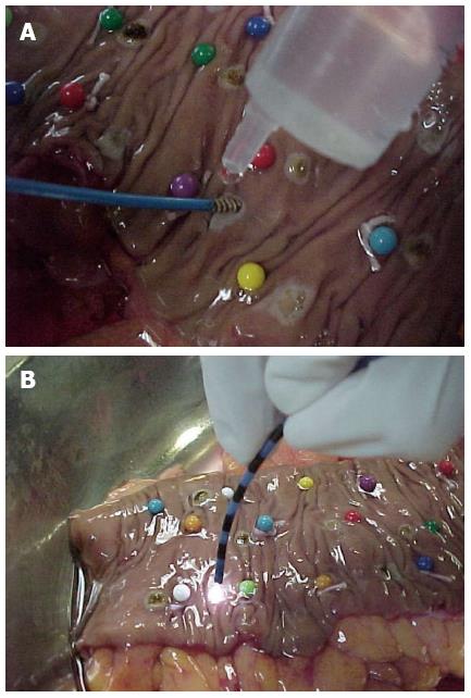
Figure 1 Colon specimen.
A: Bipolar electrocoagulation of a colon specimen; B: Argon plasma coagulation of a colon specimen.
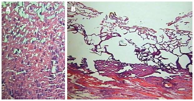
Figure 2 Cellular necrosis.
A: “Ghost cells” [hematoxylin and eosin (HE), 250 ×]; B: Citoplasmatic acidofilia and cellular picnosis (HE, 400 ×).
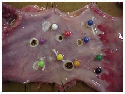
Figure 3 Macroscopic aspects of coagulated spots (both methods).
Figure 4 The microscopic aspect of coagulated spots with different depths of coagulation necrosis.
A: Microscopic aspect of superficial mucosal involvement. Citoplasmatic acidofilia, cellular picnosis in an esophageal specimen [hematoxylin and eosin (HE), 250 ×]; B: Microscopic aspect of submucosal involvement in a gastric specimen (HE, 250 ×); C: Microscopic aspect of muscularis propria involvement in a colonic specimen (HE, 250 ×).
- Citation: Garrido T, Baba ER, Wodak S, Sakai P, Cecconello I, Maluf-Filho F. Histology assessment of bipolar coagulation and argon plasma coagulation on digestive tract. World J Gastrointest Endosc 2014; 6(7): 304-311
- URL: https://www.wjgnet.com/1948-5190/full/v6/i7/304.htm
- DOI: https://dx.doi.org/10.4253/wjge.v6.i7.304


