Copyright
©2014 Baishideng Publishing Group Co.
World J Gastrointest Endosc. Feb 16, 2014; 6(2): 49-54
Published online Feb 16, 2014. doi: 10.4253/wjge.v6.i2.49
Published online Feb 16, 2014. doi: 10.4253/wjge.v6.i2.49
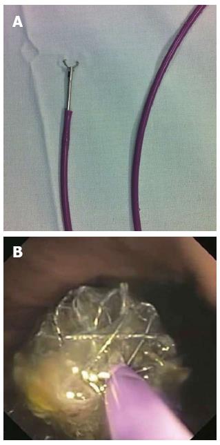
Figure 1 Endoscopic removal.
A: Rat-toothed grasper inside the biliary stent pusher; B: Stent border constrained by the grasper and pusher together.
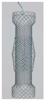
Figure 2 The Niti-S stent has a double-layered configuration.
The inner covered layer protects against tumor ingrowth and the additional uncovered mesh helps to resist migration.
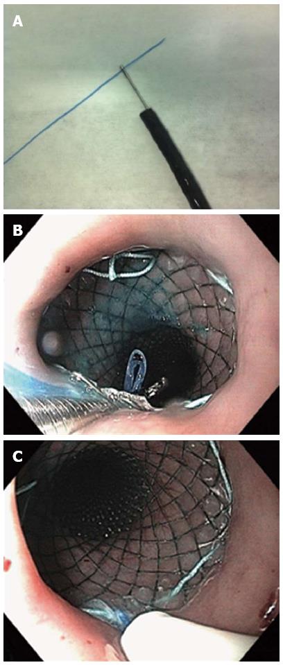
Figure 3 A length of dental floss or equivalent thread is grasped with a biopsy forceps and passed through the working channel of a standard endoscope.
A: The dental floss is grasped with a biopsy forceps; B: The dental floss is passed through the stent mesh from the outside to the inside and carried down using the biopsy forceps; C: The dental floss is inserted into a nasoenteral tube, which is pushed down towards the mesh to protect the nasopharyngeal mucosa.
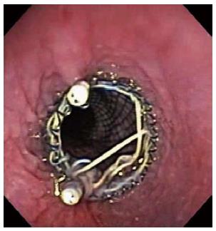
Figure 4 Endoscopic clips may be applied at the upper border of the stent to prevent migration.
- Citation: Martins BDC, Retes FA, Medrado BF, Lima MS, Pennacchi CMPS, Kawaguti FS, Safatle-Ribeiro AV, Uemura RS, Maluf-Filho F. Endoscopic management and prevention of migrated esophageal stents. World J Gastrointest Endosc 2014; 6(2): 49-54
- URL: https://www.wjgnet.com/1948-5190/full/v6/i2/49.htm
- DOI: https://dx.doi.org/10.4253/wjge.v6.i2.49


