Copyright
©2014 Baishideng Publishing Group Inc.
World J Gastrointest Endosc. Dec 16, 2014; 6(12): 630-634
Published online Dec 16, 2014. doi: 10.4253/wjge.v6.i12.630
Published online Dec 16, 2014. doi: 10.4253/wjge.v6.i12.630
Figure 1 Physical examination at admission.
The patient had a scar from the excision of hemangiomas on her right breast and multiple bluish hemangiomas on her right arm.
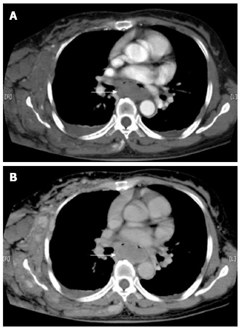
Figure 2 Dynamic computed tomography study.
A: Early phase; B: Late phase. An esophageal hematoma was found, but there was no hemoperfusion to the hematoma.
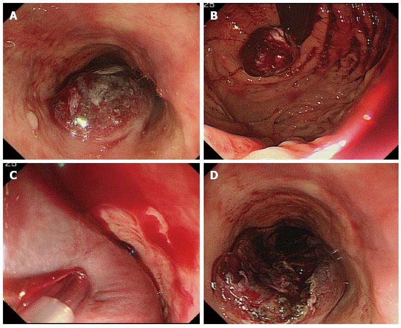
Figure 3 Endoscopic images of the esophageal hematoma taken before and during endoscopic therapy.
A and B: Esophageal hematoma from the thoracic esophagus to the gastric cardia with oozing bleeding; C: Endoscopic injection sclerotherapy with polidocanol was applied to the hematoma; D: After injection of polidocanol, the hematoma was incised using an injection needle.
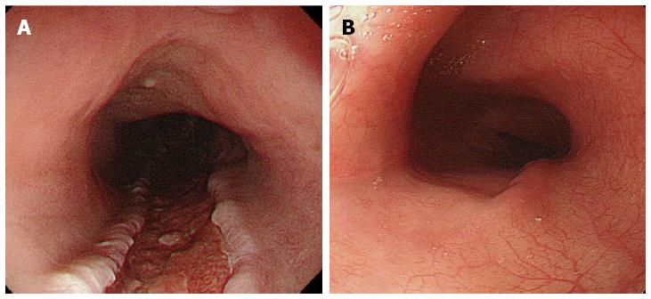
Figure 4 Endoscopic images after endoscopic therapy.
A: Seven days after endoscopic therapy, the hematoma had disappeared; B: Two months after endoscopic therapy, the esophageal ulcer healed, and the hematoma had not relapsed.
- Citation: Takasumi M, Hikichi T, Takagi T, Sato M, Suzuki R, Watanabe K, Nakamura J, Sugimoto M, Waragai Y, Kikuchi H, Konno N, Watanabe H, Obara K, Ohira H. Endoscopic therapy for esophageal hematoma with blue rubber bleb nevus syndrome. World J Gastrointest Endosc 2014; 6(12): 630-634
- URL: https://www.wjgnet.com/1948-5190/full/v6/i12/630.htm
- DOI: https://dx.doi.org/10.4253/wjge.v6.i12.630


