Copyright
©2014 Baishideng Publishing Group Inc.
World J Gastrointest Endosc. Dec 16, 2014; 6(12): 620-624
Published online Dec 16, 2014. doi: 10.4253/wjge.v6.i12.620
Published online Dec 16, 2014. doi: 10.4253/wjge.v6.i12.620
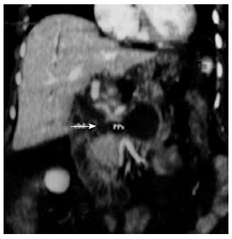
Figure 1 Coronal computed tomography reconstruction showed a large pancreatic pseudocyst with a fistulous communication (arrow) to the common bile duct.
Figure 2 Pancreatic pseudocystis intervention.
First step: Endoscopic ultrasound-guided puncture with 19-gauge dedicated needle of the pancreatic collection containing necrotic debris (A); Second step: Placement of the first plastic stent and two guidewire into the pseudocysts (PPs) for the placement of the second stent and the nasocystic drain (B); Third step: Fluoroscopic image showing the biliary stent (arrowhead), two PPs double pigtail stents and the nasocystic drain (C).
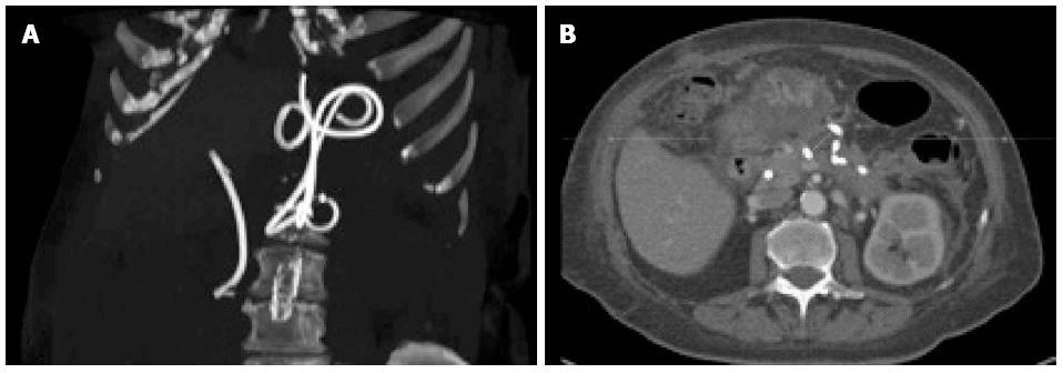
Figure 3 Computed tomography scan performed three weeks later: Scan reconstruction showing the correct position of the biliary and pancreatic collection stents (A), while the axial scan show almost complete resolution of the cystic cavity (B).
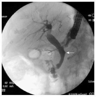
Figure 4 At endoscopic retrograde cholangiopancreatography performed 3 mo later, complete healing of the fistula was documented (arrow).
- Citation: Crinò SF, Scalisi G, Consolo P, Varvara D, Bottari A, Pantè S, Pallio S. Novel endoscopic management for pancreatic pseudocyst with fistula to the common bile duct. World J Gastrointest Endosc 2014; 6(12): 620-624
- URL: https://www.wjgnet.com/1948-5190/full/v6/i12/620.htm
- DOI: https://dx.doi.org/10.4253/wjge.v6.i12.620


