Copyright
©2014 Baishideng Publishing Group Inc.
World J Gastrointest Endosc. Dec 16, 2014; 6(12): 592-599
Published online Dec 16, 2014. doi: 10.4253/wjge.v6.i12.592
Published online Dec 16, 2014. doi: 10.4253/wjge.v6.i12.592
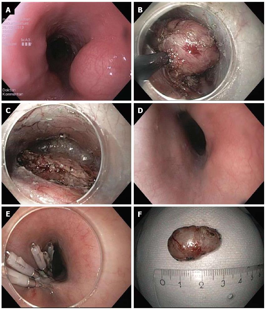
Figure 1 Submucosal endoscopic tumor resection/tunneling technique.
A: Endoscopic image of lumen obstruction subepithelial tumor in the proximal esophagus in a 42 years old woman with dysphagia; B: After preparing the submucosal tunnel, the tumor gets visible and is enucleated in endoscopic submucosal dissection-technique with a TT knife. The tumor was arising from the muscularis propria; C: Resection site (endoscope in the submucosal tunnel). The muscularis propria is excised/perforated; D: Resection site (endoscope in the esophageal lumen. Intact mucosa completely covers the muscular perforation; E: The mucosal incision (about 5 cm proximal tot he resection site) was closed with standard clips; F: Resection specimen. Histological examination revealed a Leiomyoma, which had been R0-resected.
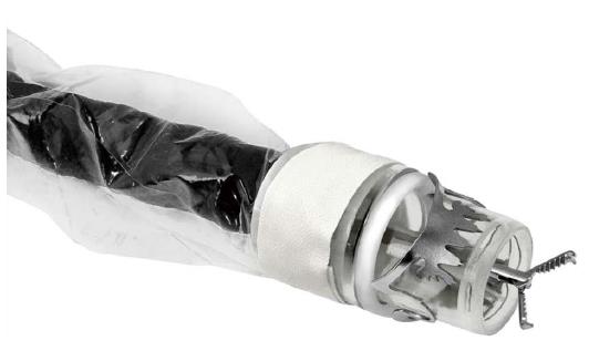
Figure 2 FTRD (Full Thickness Resection Device, Ovesco Endoscopy, Tübingen Germany).
The device is assembled on a standard colonoscope. It consists of 14 mm modified over-the-scope clips which is mounted on a long transparent cap. A monofilament snare is preloaded in the tip of the cap. The handle of the snare runs on the outer surface of the endoscope underneath a transparent sheath. A grasping forceps or a tissue anchor can be advanced through the working channel of the endoscope.
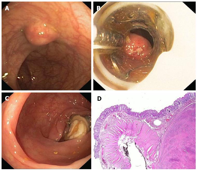
Figure 3 Endoscopic full thickness resection with the FTRD.
A: A 75 years old woman presented with a 1.5 cm subepithelial tumor in the descending colon; B: Endoscopic view with the FTRD mounted on a standard colonoscope; C: Resection site after endoscopic full thickness resection. The over-the-scope clips secures colonic wall patency; D: Histologic image (HE-staining) of the resection specimen showing one lateral resection margin. Note the cross-sectional view of the whole colonic wall on the left side. The tumor (leiomyoma) is shown on the right.
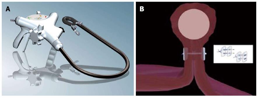
Figure 4 Endoscopic full thickness suturing.
A: The GERDX suturing device (G-Surg, Seeon, Germany); B: Schematic illustration of full thickness suturing. Application of PTFE-pledgeted sutures underneath the tumor creates a gastric wall duplication with serosa-to-serosa apposition.
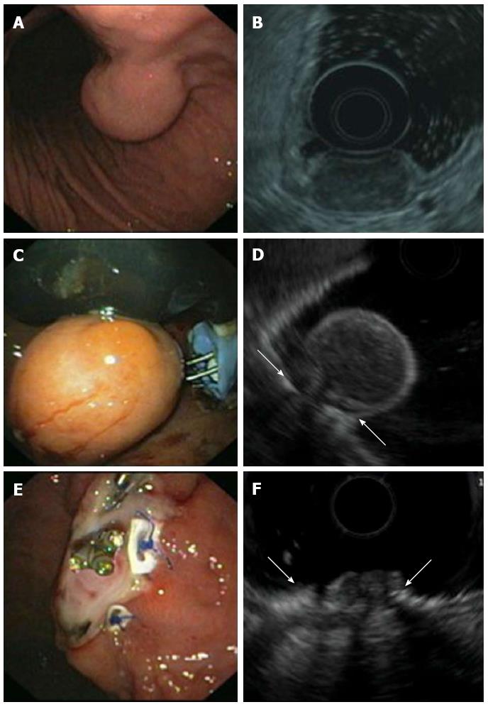
Figure 5 Endoscopic full thickness resection of gastric gastrointestinal stromal tumors after transmural suturing.
A: Subepithelial tumor in the gastric corpus; B: Endoscopic ultrasound (EUS) showed a hypoechoic tumor originating from the muscularis propria with a maximum diameter of 27 mm; C: Two transmural sutures underneath the tumor were applied using the PlicatorTM suturing device; D: EUS image of the pseudopolyp after suturing. Arrows are indicating the sutures; E: The tumor was resected with a snare above the sutures. The transmural PTFE-pledgeted sutures are securing gastric wall patency. Resection was macroscopically complete; F: EUS image of the resection site. Arrows are indicating the sutures. There was no evidence of residual tumor.
- Citation: Schmidt A, Bauder M, Riecken B, Caca K. Endoscopic resection of subepithelial tumors. World J Gastrointest Endosc 2014; 6(12): 592-599
- URL: https://www.wjgnet.com/1948-5190/full/v6/i12/592.htm
- DOI: https://dx.doi.org/10.4253/wjge.v6.i12.592


