Copyright
©2014 Baishideng Publishing Group Inc.
World J Gastrointest Endosc. Nov 16, 2014; 6(11): 534-540
Published online Nov 16, 2014. doi: 10.4253/wjge.v6.i11.534
Published online Nov 16, 2014. doi: 10.4253/wjge.v6.i11.534
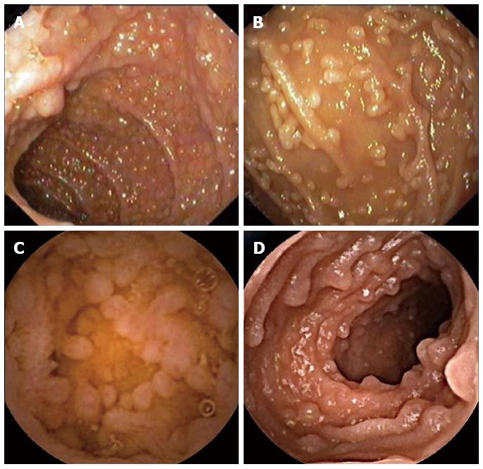
Figure 1 Endoscopic appearance of nodular lymphoid hyperplasia.
A: Upper endoscopy revealing a nodular lymphoid hyperplasia of the duodenal bulb in a patient with common variable immunodeficiency; B: Upper endoscopy showing nodular lymphoid hyperplasia of the second part of the duodenum in a patient with selective IgA deficiency; C: Small bowel capsule endoscopy revealing a nodular lymphoid hyperplasia of the jejunum in a patient selective IgA deficiency; D: Small bowel capsule endoscopy revealing a nodular lymphoid hyperplasia of the terminal ileum in a patient with common variable immunodeficiency.
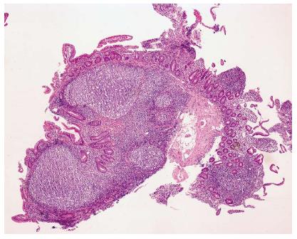
Figure 2 Duodenal biopsies stained with haematoxylin-eosin (× 40), with hyperplasic lymphoid follicles, suggesting nodular lymphoid hyperplasia.
- Citation: Albuquerque A. Nodular lymphoid hyperplasia in the gastrointestinal tract in adult patients: A review. World J Gastrointest Endosc 2014; 6(11): 534-540
- URL: https://www.wjgnet.com/1948-5190/full/v6/i11/534.htm
- DOI: https://dx.doi.org/10.4253/wjge.v6.i11.534


