Copyright
©2014 Baishideng Publishing Group Inc.
World J Gastrointest Endosc. Oct 16, 2014; 6(10): 506-509
Published online Oct 16, 2014. doi: 10.4253/wjge.v6.i10.506
Published online Oct 16, 2014. doi: 10.4253/wjge.v6.i10.506
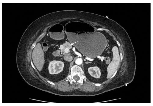
Figure 1 Computer tomographic of the abdomen and pelvis with pancreas protocol showed a 1.
7 cm × 1.2 cm × 1.6 cm solid mass in the superior and right lateral margin of the head pancreas. The mass was hyperdense to the pancreas on early arterial phase imaging and became isodense with washout on more delayed phase images.
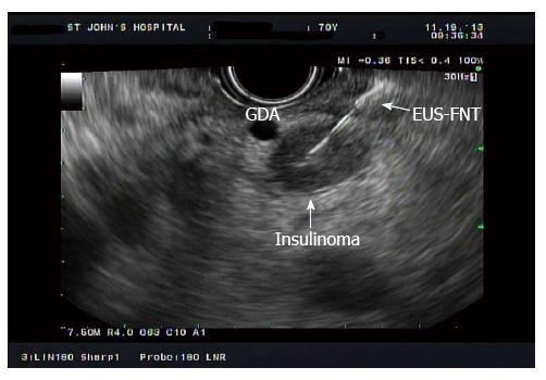
Figure 2 Endoscopic ultrasound-guided fine- needle tattooing image of a 15.
5-mm hypoechoic mass located at pancreatic head adjacent to gastroduodenal artery.
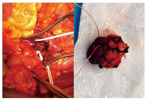
Figure 3 The tattooed insulinoma, located between gastroduodenal artery and superior mesenteric vein, was identified during operation (left).
The 2 cm Insulinoma was enucleated without complication (right).
- Citation: Leelasinjaroen P, Manatsathit W, Berri R, Barawi M, Gress FG. Role of preoperative endoscopic ultrasound-guided fine-needle tattooing of a pancreatic head insulinoma. World J Gastrointest Endosc 2014; 6(10): 506-509
- URL: https://www.wjgnet.com/1948-5190/full/v6/i10/506.htm
- DOI: https://dx.doi.org/10.4253/wjge.v6.i10.506


