Copyright
©2014 Baishideng Publishing Group Co.
World J Gastrointest Endosc. Jan 16, 2014; 6(1): 27-31
Published online Jan 16, 2014. doi: 10.4253/wjge.v6.i1.27
Published online Jan 16, 2014. doi: 10.4253/wjge.v6.i1.27
Figure 1 Positive manifestations in examination and in the surgically resected segment.
A: A giant and ovoid ulceration in the inferior extremity of the esophagus; B: A typical oval-shaped large ulcer at the ileocecal junction; C: A few oval ulcers around the crissum; D: The resected material showed that the wall of the cecum was thick, occasional discoloration in the serosa, mucosal edema, an ulcer (4 cm × 4 cm) and occasional necrosis in a segment 32 cm in length involving the ileocecal region.
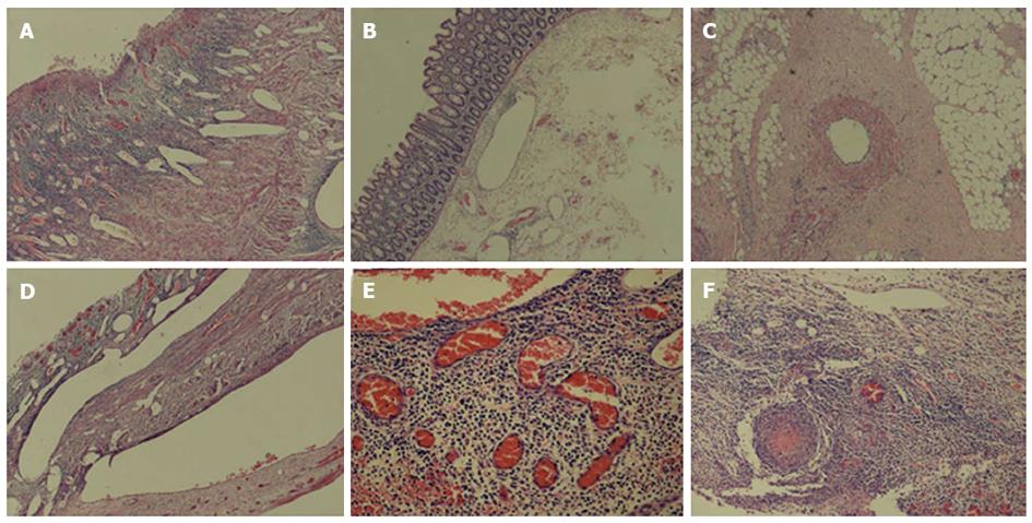
Figure 2 Positive pathological manifestations in the surgically resected segment.
A: The ulcer in the ileocecal lesion encroached the whole segment with ectasia and blood vessel hyperplasia [hematoxylin and eosin (HE), × 40]; B: Ectasis in blood vessels was observed in normal tissues around the lesion (HE, × 40); C: The thickened vessel intima with lymphocyte and polymorphonuclear leukocyte infiltration (HE, × 40); D: Extreme ectasia in lesion blood vessels (HE, × 40); E: There was considerable lymphocyte infiltration in and around the vessel wall (HE, × 200); F: Thrombus and recanalization in some vessels at the base of the ulcer (HE, × 100).
- Citation: Wang ZK, Shi H, Wang SD, Liu J, Zhu WM, Yang MF, Liu C, Lu H, Wang FY. Confusing untypical intestinal Behcet’s disease: Skip ulcers with severe lower gastrointestinal hemorrhage. World J Gastrointest Endosc 2014; 6(1): 27-31
- URL: https://www.wjgnet.com/1948-5190/full/v6/i1/27.htm
- DOI: https://dx.doi.org/10.4253/wjge.v6.i1.27


