Copyright
©2014 Baishideng Publishing Group Co.
World J Gastrointest Endosc. Jan 16, 2014; 6(1): 13-19
Published online Jan 16, 2014. doi: 10.4253/wjge.v6.i1.13
Published online Jan 16, 2014. doi: 10.4253/wjge.v6.i1.13
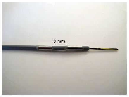
Figure 1 The radiofrequency ablation probe Habib EndoHPB (EMcision United Kingdom, London, United Kingdom) features two ring electrodes at the tip that are 8 mm apart.
The probe is designed to perform bipolar cautery in endoscopic surgical procedures.
Figure 2 Exemplary results from the ex vivo pig liver model.
From left to right, higher watt variables were used. Necrotic areas are marked by a arrow.
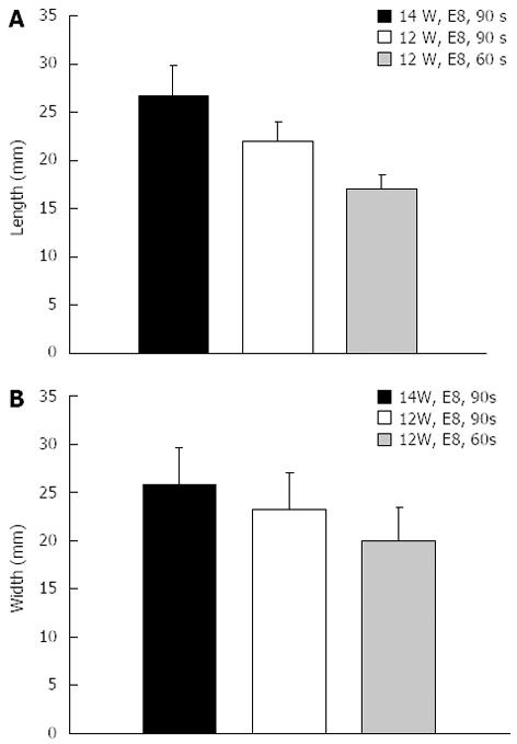
Figure 3 Results from the ex-vivo pig liver model.
A: Length; B: Width. Varying electrosurgical variables revealed distinct differences in the extent of necrotic area. The used combinations are explained in the legend. e.g., the blue column shows length and width of the necrosis caused by RFA with 14 watt, effect 8 with an ablation time of 90 s. RFA: Radiofrequency ablation.
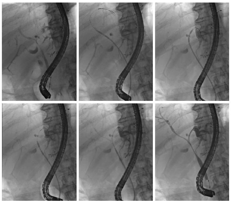
Figure 4 Application of endoscopically guided, intraductal radiofrequency ablation in a 72-year-old patient with an extended perihilar cholangiocarcinoma (Klatskin tumor, stage Bismuth IV, histologically proven) involving all subsegments.
Multisegmental radiofrequency ablation (RFA) applications were performed (from left above to lower right). The patient experienced no treatment-associated complications and was doing well 15 mo after the initiation of endoscopic RFA treatment.
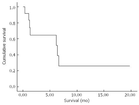
Figure 5 Kaplan-Meier survival curve of all study patients (n = 12).
Calculation of survival started at the time of the first endoscopic radiofrequency ablation treatment in each patient.
- Citation: Tal AO, Vermehren J, Friedrich-Rust M, Bojunga J, Sarrazin C, Zeuzem S, Trojan J, Albert JG. Intraductal endoscopic radiofrequency ablation for the treatment of hilar non-resectable malignant bile duct obstruction. World J Gastrointest Endosc 2014; 6(1): 13-19
- URL: https://www.wjgnet.com/1948-5190/full/v6/i1/13.htm
- DOI: https://dx.doi.org/10.4253/wjge.v6.i1.13


