Copyright
©2013 Baishideng Publishing Group Co.
World J Gastrointest Endosc. Jun 16, 2013; 5(6): 293-296
Published online Jun 16, 2013. doi: 10.4253/wjge.v5.i6.293
Published online Jun 16, 2013. doi: 10.4253/wjge.v5.i6.293
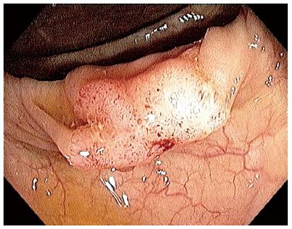
Figure 1 Endoscopic image showing a polypoid lesion in the transverse colon.
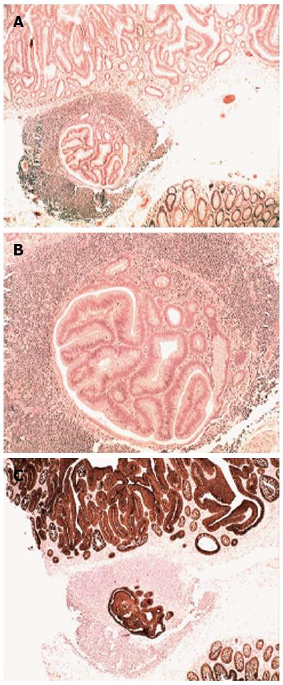
Figure 2 Low-power view.
A: A villous adenoma on top of a gut-associated lymphoid tissue (GALT) with carcinoma [hematoxylin and eosin (HE) × 4]; B: Detail showing carcinoma in GALT (HE × 10); C: A villous adenoma on top of a GALT with carcinoma (MNF 116 × 4).
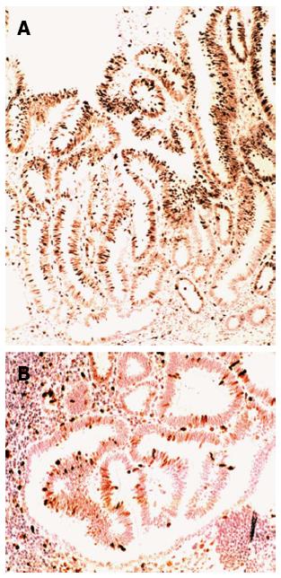
Figure 3 High-power view.
A: The villous adenoma showing high cell proliferation (Ki67, clone MIB1 × 10); B: Gut-associated lymphoid tissue with carcinoma showing lower cell proliferation than in the villous adenoma on top (Ki67, clone MIB1 × 20).
- Citation: Rubio CA, Befrits R, Ericsson J. Carcinoma in gut-associated lymphoid tissue in ulcerative colitis: Case report and review of literature. World J Gastrointest Endosc 2013; 5(6): 293-296
- URL: https://www.wjgnet.com/1948-5190/full/v5/i6/293.htm
- DOI: https://dx.doi.org/10.4253/wjge.v5.i6.293


