Copyright
©2013 Baishideng Publishing Group Co.
World J Gastrointest Endosc. Jun 16, 2013; 5(6): 275-280
Published online Jun 16, 2013. doi: 10.4253/wjge.v5.i6.275
Published online Jun 16, 2013. doi: 10.4253/wjge.v5.i6.275
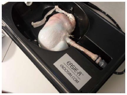
Figure 1 Simulation platform using the EASIE-R simulator with an ex-vivo porcine stomach specimen.
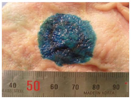
Figure 2 3 cm × 3 cm target lesions created by methylene blue tattoo in the mucosa of fresh ex-vivo stomachs.
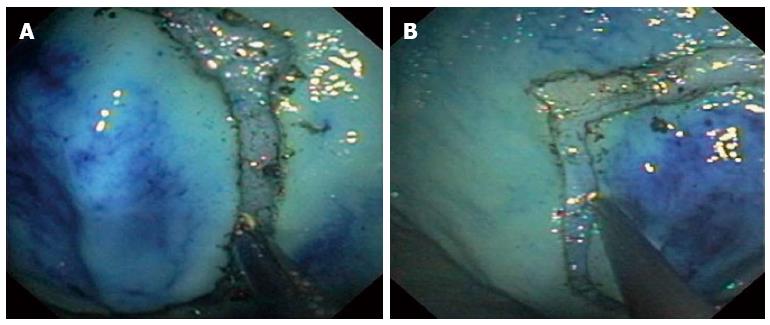
Figure 3 Endoscopic images.
A: Circumferential resection with the IT knife after injection; B: The separation of the circumferential cutting area being carefully inspected.
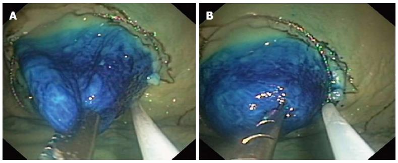
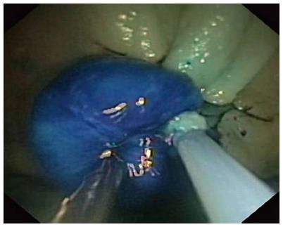
Figure 4 Endoscopic images.
A: The mucosal retraction with regular forceps (unipolar traction); B: Mucosal retraction with the tissue-anchoring device (retracting tissue from three anchor points).
Figure 5 The snare being subsequently closed and the specimen resected by application of electrocautery.
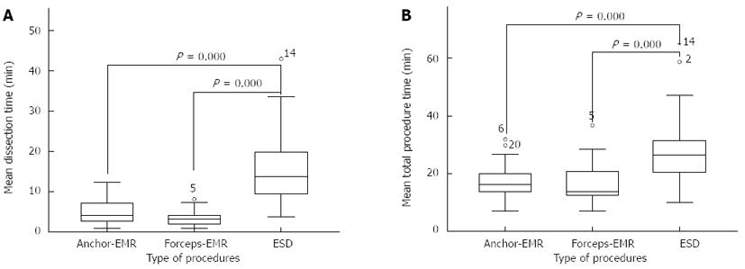
Figure 6 The overall mean total procedure time.
A: Overall mean dissection time; B: Mean total procedure time. EMR: Endoscopic mucosal resection; ESD: Endoscopic submucosal dissection.
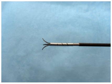
Figure 7 Detail of the novel tissue-anchoring device.
- Citation: Jung Y, Kato M, Lee J, Gromski MA, Chuttani R, Matthes K. Effectiveness of circumferential endoscopic mucosal resection with a novel tissue-anchoring device. World J Gastrointest Endosc 2013; 5(6): 275-280
- URL: https://www.wjgnet.com/1948-5190/full/v5/i6/275.htm
- DOI: https://dx.doi.org/10.4253/wjge.v5.i6.275


