Copyright
©2012 Baishideng Publishing Group Co.
World J Gastrointest Endosc. Jul 16, 2012; 4(7): 335-338
Published online Jul 16, 2012. doi: 10.4253/wjge.v4.i7.335
Published online Jul 16, 2012. doi: 10.4253/wjge.v4.i7.335
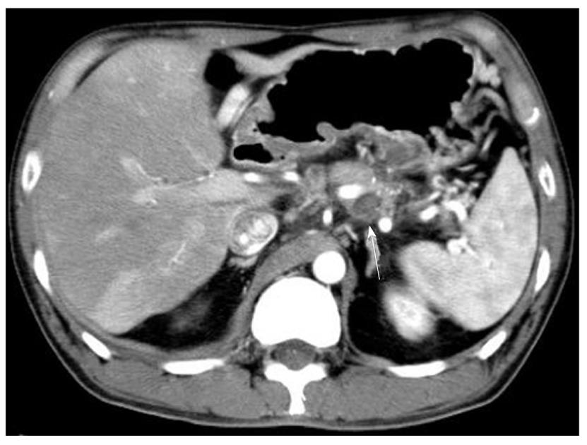
Figure 1 Abdominal computed tomographic findings.
A severely atrophied pancreas with a calcified body was noted. The pseudocyst (arrow) ranged from the back of the pancreas to the left mediastinum and was adjacent to the splenic artery.
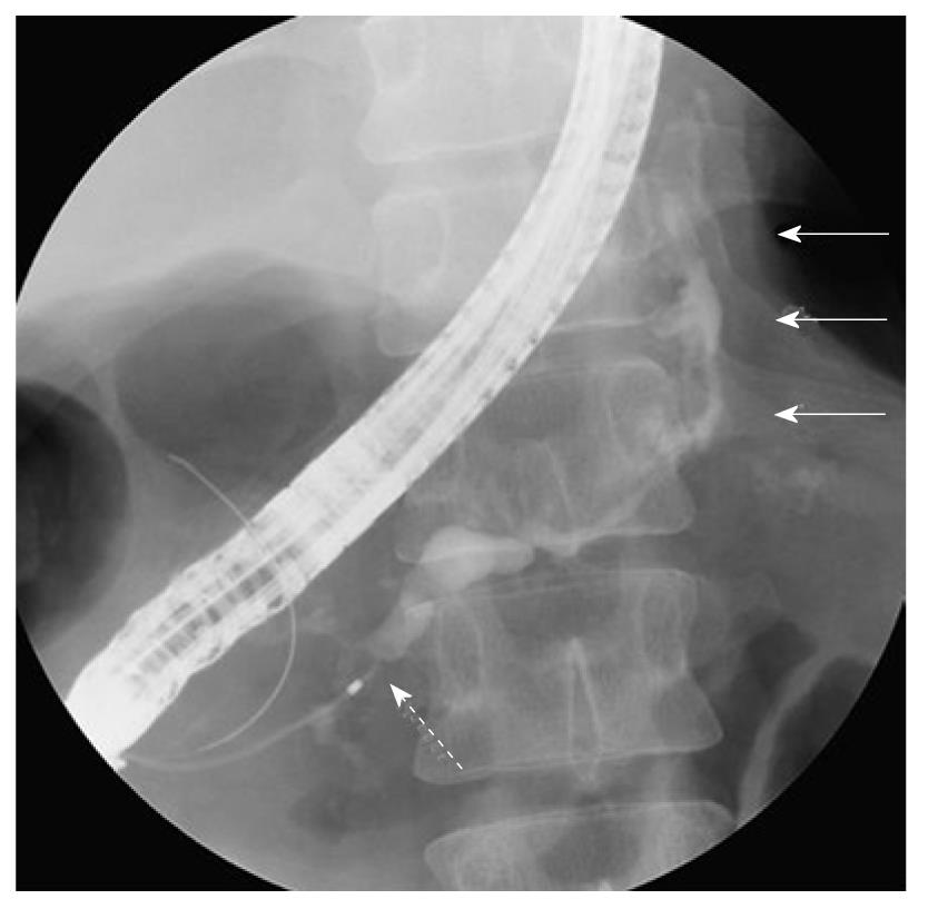
Figure 2 Endoscopic retrograde pancreatography findings.
A: Endoscopic retrograde pancreatography showed stenosis of the principal pancreatic duct at the pancreatic head (dotted arrow) and a dilated tail duct communicating with the left mediastinal pseudocyst (solid arrows).
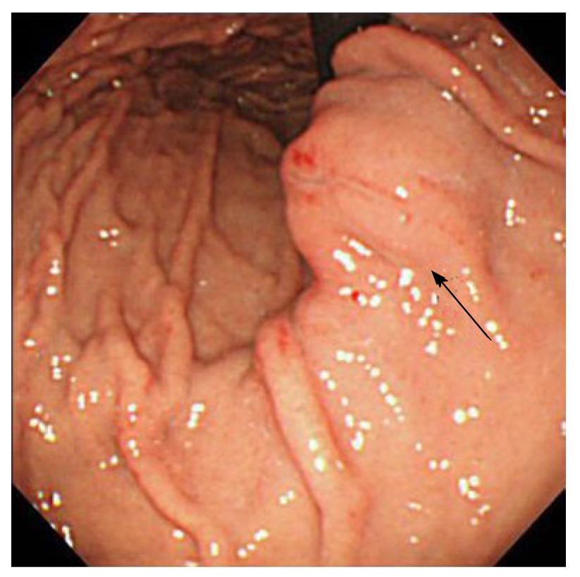
Figure 3 Upper gastrointestinal endoscopy findings.
Upper gastrointestinal endoscopy showed a 2 cm, submucosal tumor-like protrusion with a red, eroded upper region located in the lesser curvature of the middle of the body of the stomach (arrow).
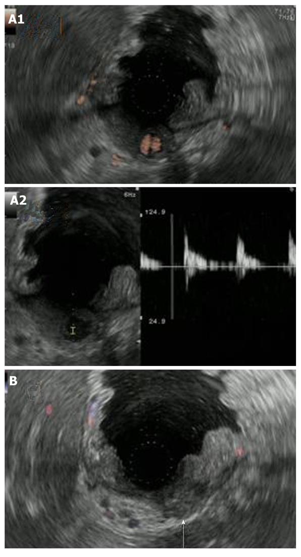
Figure 4 Endoscopic ultrasonography findings.
A1: Endoscopic ultrasonography showed an anechoic region whose entire periphery was hypoechoic beneath the gastric mucosa. Power Doppler showed blood flow in the anechoic region. Upper gastrointestinal endoscopy showed a 2 cm, submucosal tumor-like protrusion with a red, eroded upper region located in the lesser curvature of middle of the body of the stomach (arrow); A2: Pulsed wave Doppler showed pulsatile blood flow in the anechoic region. This finding led to the diagnosis of an aneurysm; B: The cessation of blood flow to the pseudoaneurysm was confirmed with endoscopic ultrasonography which was performed 1 wk after treatment (arrow).
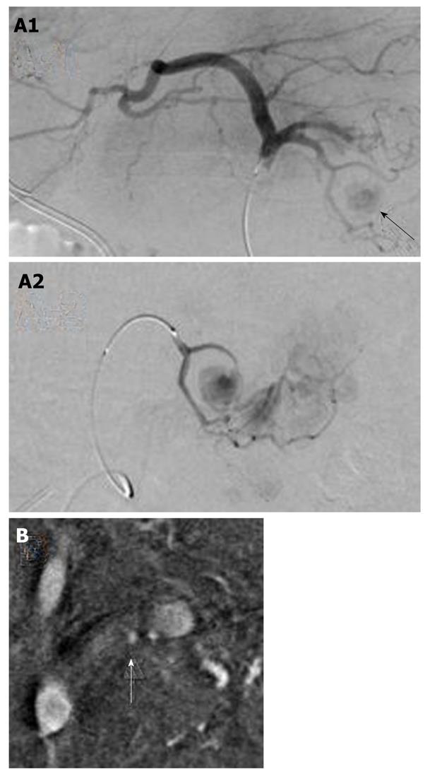
Figure 5 Angiography findings.
A1: The pseudoaneurysm of the left gastric artery was diagnosed on angiography (arrow). The left hepatic artery diverged from the left gastric artery; A2: The microcatheter was advanced in the region of the pseudoaneurysm, and the pseudoaneurysm was embolized with histoacryl and lipiodol; B: A small pseudoaneurysm was observed in the splenic artery (arrow), and the splenic artery was embolized by coils.
- Citation: Fukatsu K, Ueda K, Maeda H, Yamashita Y, Itonaga M, Mori Y, Moribata K, Shingaki N, Deguchi H, Enomoto S, Inoue I, Maekita T, Iguchi M, Tamai H, Kato J, Ichinose M. A case of chronic pancreatitis in which endoscopic ultrasonography was effective in the diagnosis of a pseudoaneurysm. World J Gastrointest Endosc 2012; 4(7): 335-338
- URL: https://www.wjgnet.com/1948-5190/full/v4/i7/335.htm
- DOI: https://dx.doi.org/10.4253/wjge.v4.i7.335


