Copyright
©2012 Baishideng Publishing Group Co.
World J Gastrointest Endosc. Jul 16, 2012; 4(7): 328-330
Published online Jul 16, 2012. doi: 10.4253/wjge.v4.i7.328
Published online Jul 16, 2012. doi: 10.4253/wjge.v4.i7.328
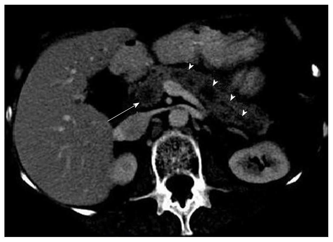
Figure 1 Computed tomography scan of the abdomen showing multiple cystic lesions throughout the pancreas (arrow heads), the largest seen in the pancreatic head (arrow).
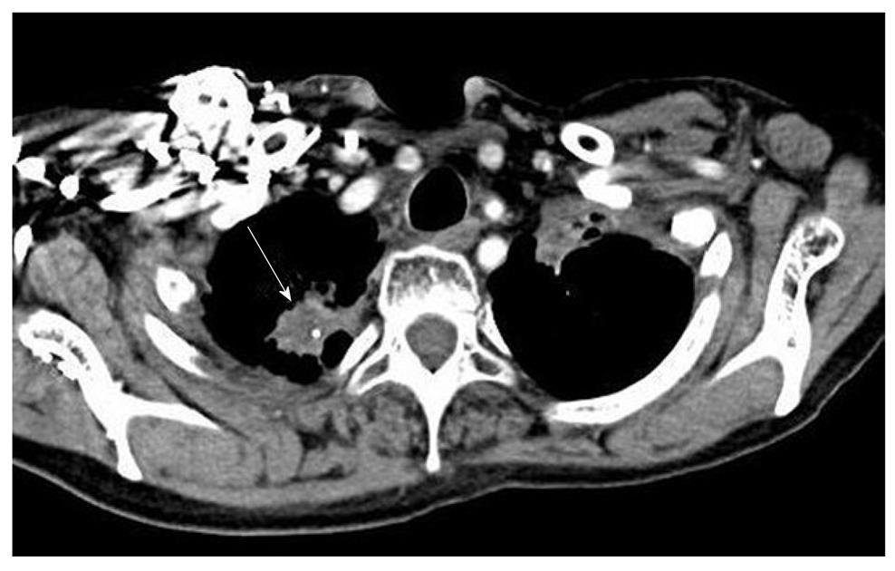
Figure 2 Computed tomography scan of the chest demonstrating the spiculated right apical lesion (arrow) with size and characteristics similar to the previously diagnosed small-cell lung cancer.
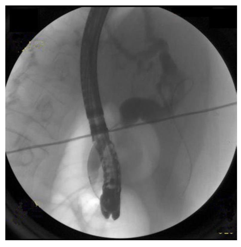
Figure 3 Endoscopic retrograde cholangiopancreatography showing dilated ducts.
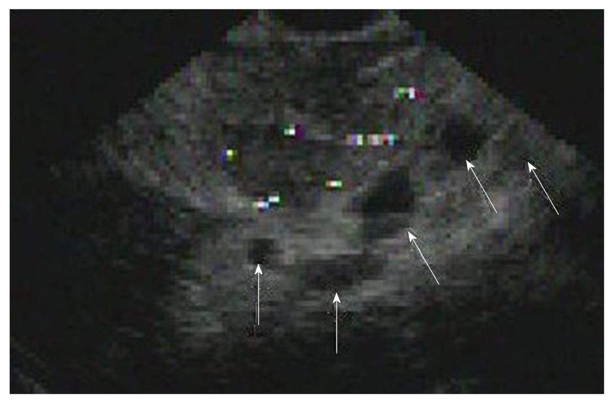
Figure 4 Endoscopic ultrasound image depicting multiple hypo-echoic lesions throughout the pancreas (arrows).
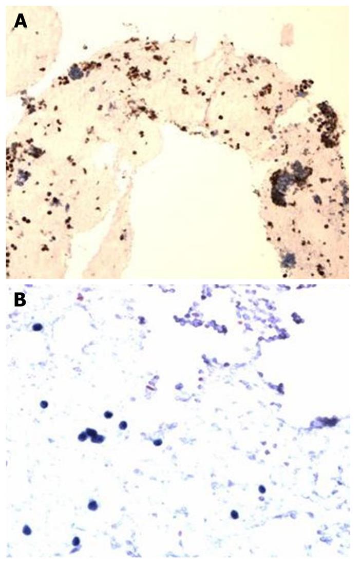
Figure 5 Fine needle aspiration cytology of the pancreas showing (A) quick stain of pancreatic mass showing small round blue cells, resembling small cell tumor, and (B) cell block of the pancreatic tissue with tumor cells positive for TTF-1 and chromogranin, indicating the origin of the tumor (lung).
- Citation: Singh D, Vaidya OU, Sadeddin E, Yousef O. Role of endoscopic ultrasound and endoscopic retrograde cholangiopancreatography in isolated pancreatic metastasis from lung cancer. World J Gastrointest Endosc 2012; 4(7): 328-330
- URL: https://www.wjgnet.com/1948-5190/full/v4/i7/328.htm
- DOI: https://dx.doi.org/10.4253/wjge.v4.i7.328


