Copyright
©2012 Baishideng Publishing Group Co.
World J Gastrointest Endosc. Jun 16, 2012; 4(6): 241-246
Published online Jun 16, 2012. doi: 10.4253/wjge.v4.i6.241
Published online Jun 16, 2012. doi: 10.4253/wjge.v4.i6.241
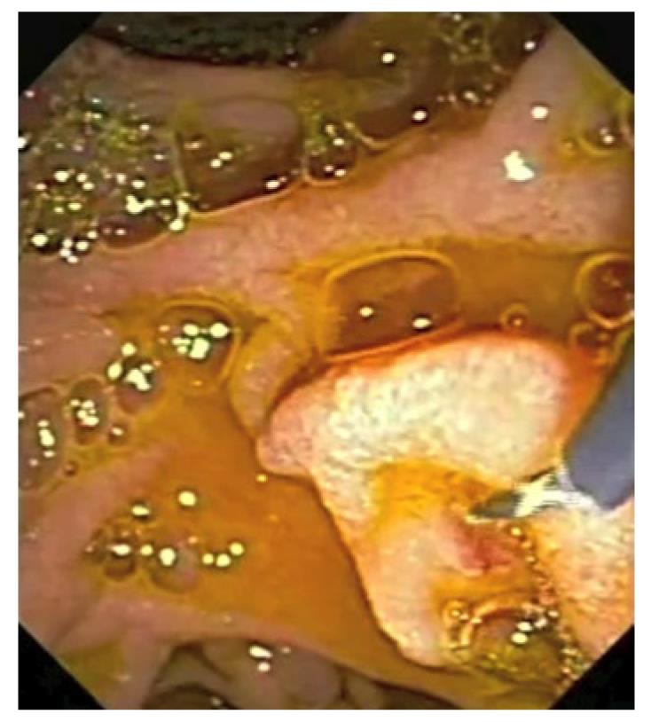
Figure 1 The guide-wire technique has been used in this patient to cannulate the minor papilla.
The minor papilla is cannulated with the guide-wire tip protruding a few millimeters over the cannula.
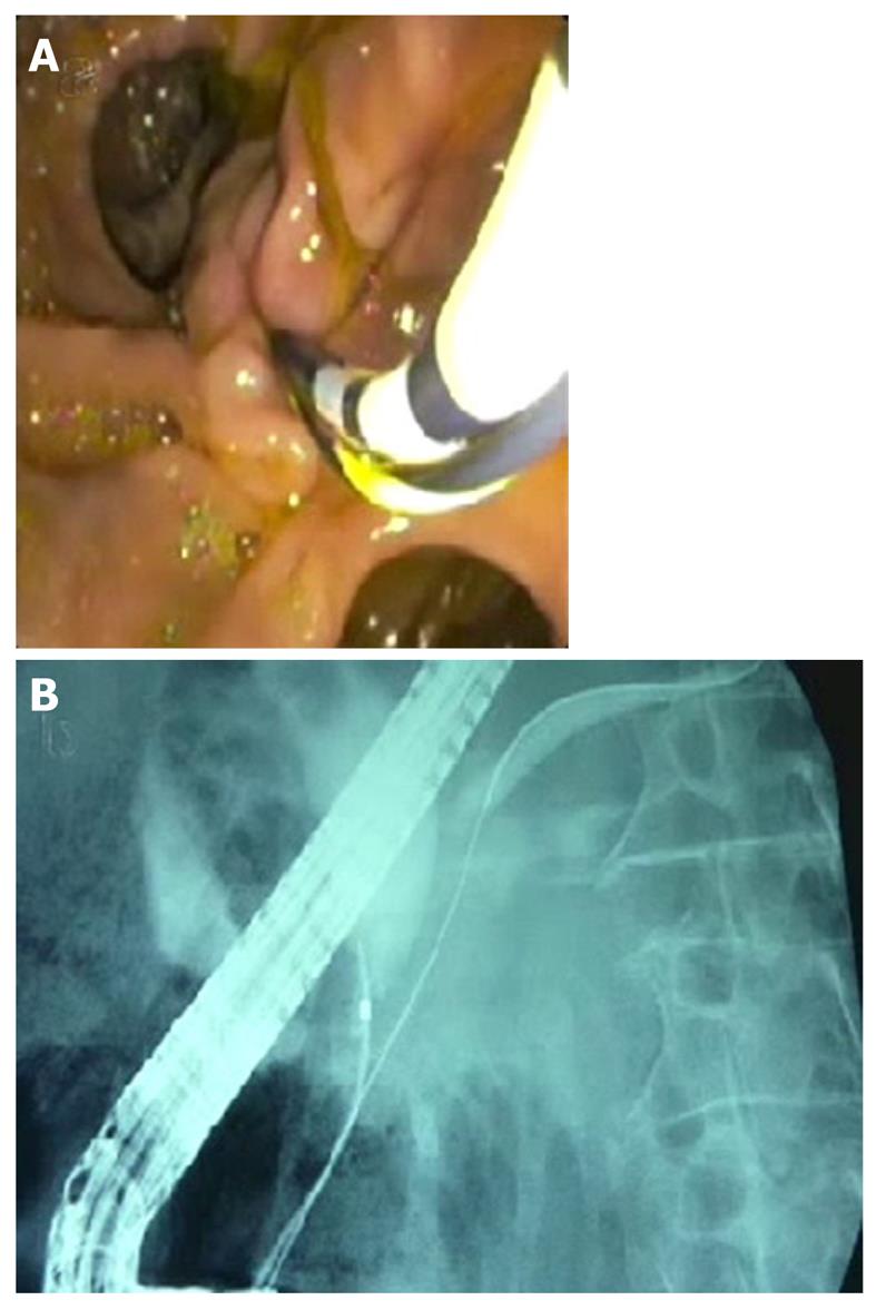
Figure 2 Image of the double guide-wire technique.
A guide-wire is inserted in the pancreatic duct and left in situ. The cannula is then inserted parallel to the pancreatic guide-wire (A) in order to cannulate the bile duct (B).
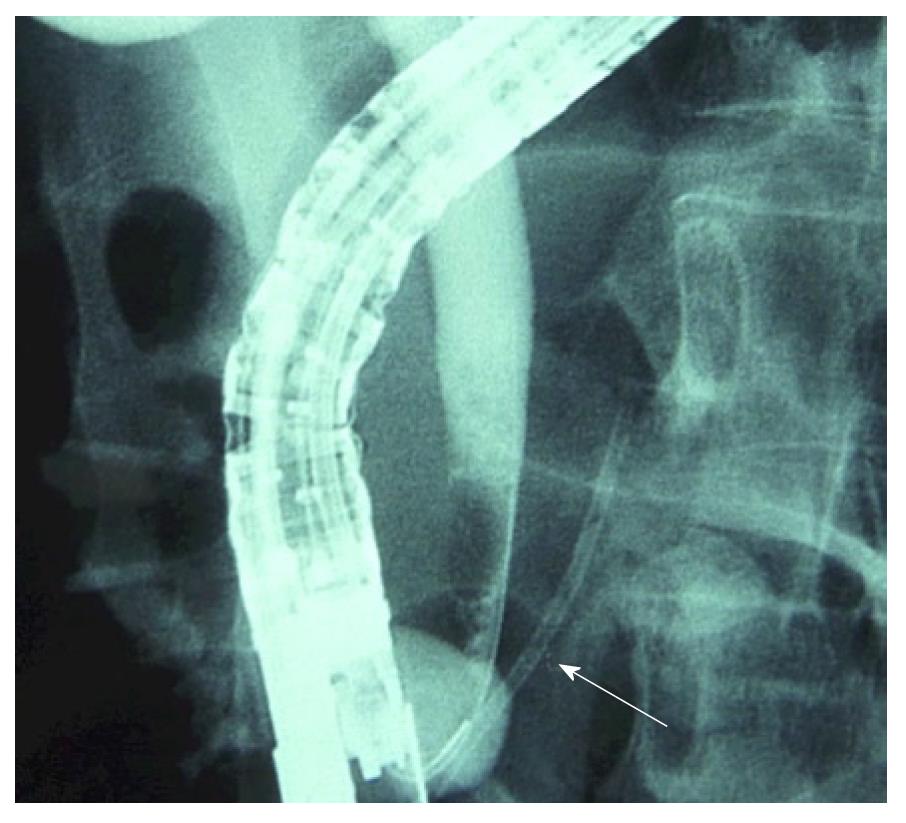
Figure 3 In this fluoroscopic image biliary cannulation over a pancreatic stent technique is shown.
The technique resembles the double-wire technique, but a plastic stent (arrow) is inserted in the pancreatic duct instead of the guide-wire, and the bile duct is cannulated in parallel.
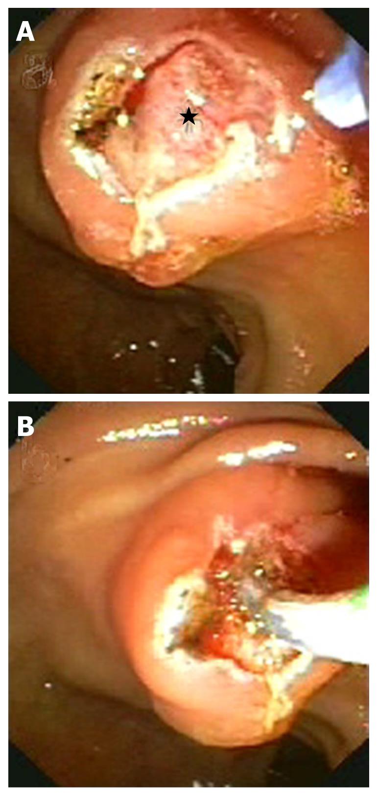
Figure 4 Needle knife sphincterotomy technique.
A superficial mucosal cut is performed showing the duodenal portion of the common bile duct as a reddish rounded protrusion (asterisk) (A). A deeper cut is made on this nodule going into the bile duct (B).
- Citation: Vila JJ, Artifon EL, Otoch JP. Post-endoscopic retrograde cholangiopancreatography complications: How can they be avoided? World J Gastrointest Endosc 2012; 4(6): 241-246
- URL: https://www.wjgnet.com/1948-5190/full/v4/i6/241.htm
- DOI: https://dx.doi.org/10.4253/wjge.v4.i6.241


