Copyright
©2012 Baishideng Publishing Group Co.
World J Gastrointest Endosc. May 16, 2012; 4(5): 162-166
Published online May 16, 2012. doi: 10.4253/wjge.v4.i5.162
Published online May 16, 2012. doi: 10.4253/wjge.v4.i5.162
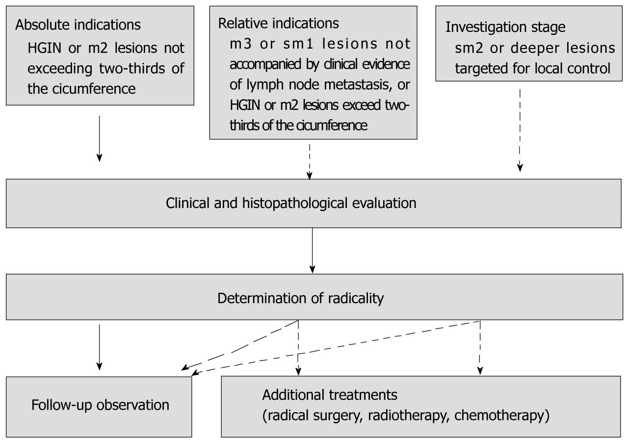
Figure 1 Indication for endoscopic resection in the Japan Esophageal Society guideline.
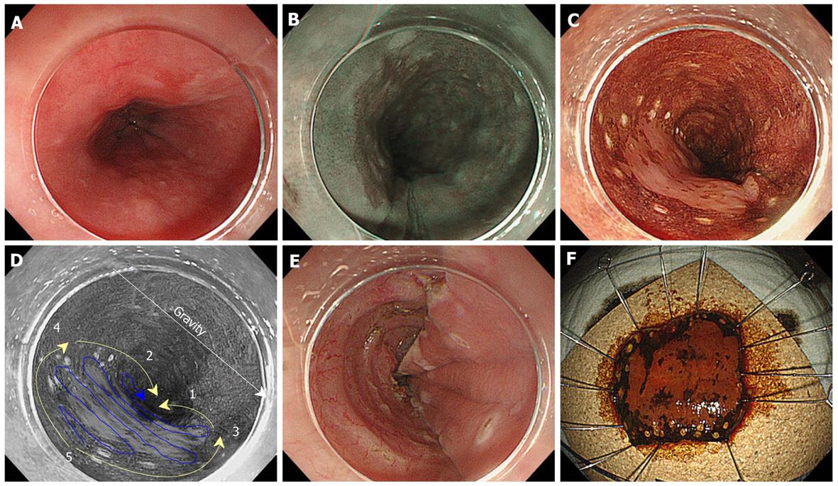
Figure 2 Endoscopic submucosal dissection of an esophageal neoplasm.
A: The reddish mucosa in the anterior wall of the middle thoracic esophagus shown by conventional endoscopy with white light; B: The brownish mucosa in one-third of the circumferential extension shown by endoscopy with narrow band imaging; C: Marking around the lesion under chromoendoscopy with iodine staining to demarcate the lesion; D: Mucosal incision at the anal side (yellow line 1-2), followed by incision at the oral side (yellow line 3-4). Incision is made from the lower side to lift it up from the collection of fluid taking gravity into consideration. After circumferential incision, dissection of the submucosa is begun from the oral end to the anal end (blue line 5); E: Artificial ulcer after removal of the lesion; F: Resected specimen in an en bloc fashion.
- Citation: Ono S, Fujishiro M, Koike K. Endoscopic submucosal dissection for superficial esophageal neoplasms. World J Gastrointest Endosc 2012; 4(5): 162-166
- URL: https://www.wjgnet.com/1948-5190/full/v4/i5/162.htm
- DOI: https://dx.doi.org/10.4253/wjge.v4.i5.162


