Copyright
©2012 Baishideng Publishing Group Co.
World J Gastrointest Endosc. Dec 16, 2012; 4(12): 575-578
Published online Dec 16, 2012. doi: 10.4253/wjge.v4.i12.575
Published online Dec 16, 2012. doi: 10.4253/wjge.v4.i12.575
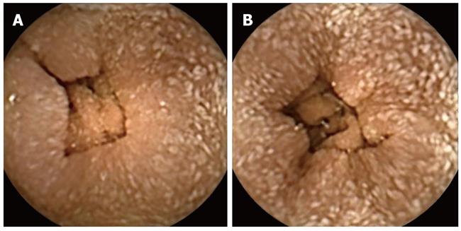
Figure 1 Video capsule examination showing small, white, diffuse deposits, covering all the small bowel mucosa (A and B).
The villi are present and edematous.
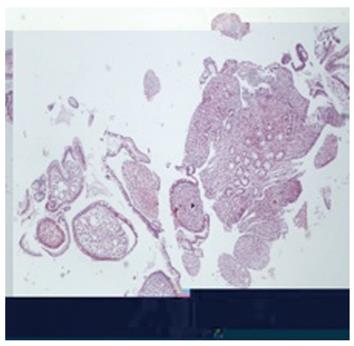
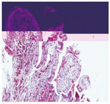
Figure 2 Whipple’s disease, small intestine with flattened villi, expanded by a dense infiltrate of foamy macrophages (hematoxylin and eosin, 4 x).
Mall bowel mucosa; the villi are present and edematous.
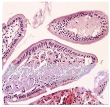
Figure 3 Whipple’s disease-small intestine with polymorphonuclear and foamy macrophages (hematoxylin and eosin, 20 x).
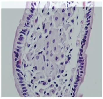
Figure 4 Whipple’s disease-intestinal villi with foamy macrophages (hematoxylin and eosin, 40 x).
Figure 5 Whipple’s disease-foamy macrophages with Periodic Acid Schiff positive bacilli in the cytoplasm (10 x).
- Citation: Mateescu BR, Bengus A, Marinescu M, Staniceanu F, Micu G, Negreanu L. First Pillcam Colon 2 capsule images of Whipple’s disease: Case report and review of the literature. World J Gastrointest Endosc 2012; 4(12): 575-578
- URL: https://www.wjgnet.com/1948-5190/full/v4/i12/575.htm
- DOI: https://dx.doi.org/10.4253/wjge.v4.i12.575


