Copyright
©2012 Baishideng Publishing Group Co.
World J Gastrointest Endosc. Dec 16, 2012; 4(12): 545-555
Published online Dec 16, 2012. doi: 10.4253/wjge.v4.i12.545
Published online Dec 16, 2012. doi: 10.4253/wjge.v4.i12.545
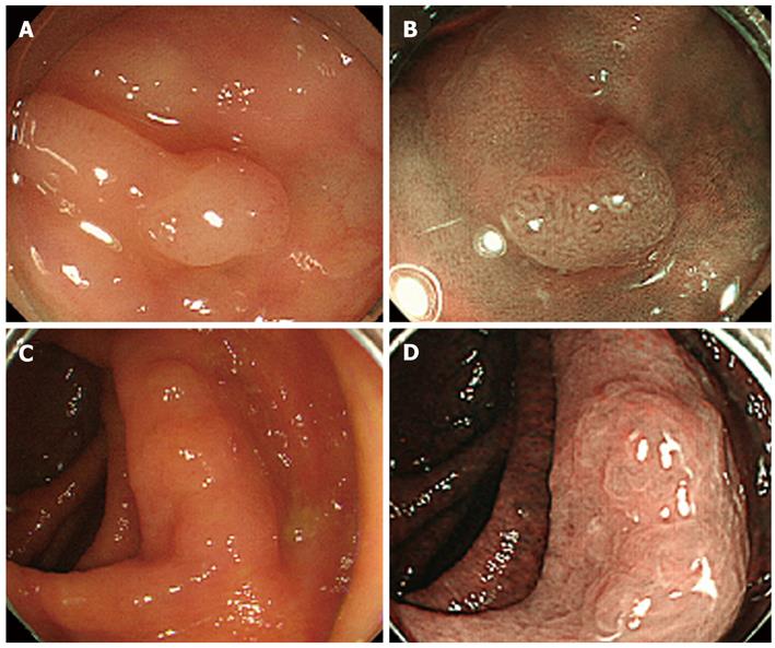
Figure 1 Narrow band imaging without magnification.
A: Ia polyp 3 mm in diameter. White-light (WI) endoscopy figure; B: Meshed capillary pattern and oval surface pattern were detected with narrow band imaging (NBI) without magnification. The polyp was diagnosed as a neoplastic polyp; C: IIa polyp 16 mm in diameter. WI endoscopy figure; D: Meshed capillary pattern was not detected with NBI without magnification, but a round surface pattern was detected. The polyp was diagnosed as a non-neoplastic polyp.
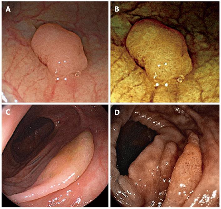
Figure 2 Flexible spectral imaging color enhancement without magnification.
A: Ia polyp 5 mm in diameter. White-light (WI) endoscopy figure; B: Flexible spectral imaging color enhancement (FICE) without magnification. FICE settings were RGB wavelengths 550, 500 and 470 nm. Round pits were identified as non-neoplastic surface patterns; C: IIa polyp 16 mm in diameter. WI endoscopy figure; D: Meshed capillary pattern was not detected with FICE without magnification, but round surface pattern was detected. The polyp was diagnosed as a non-neoplastic polyp.
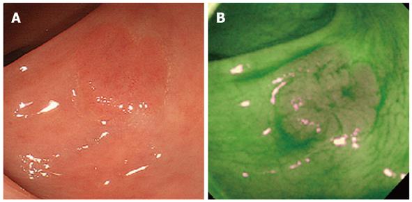
Figure 3 Autofluorescence imaging.
A: IIa polyp 14 mm in diameter (White-light endoscopy figure); B: In autofluorescence imaging, the normal mucosa was detected by green color and the neoplastic polyp was detected by magenta color.
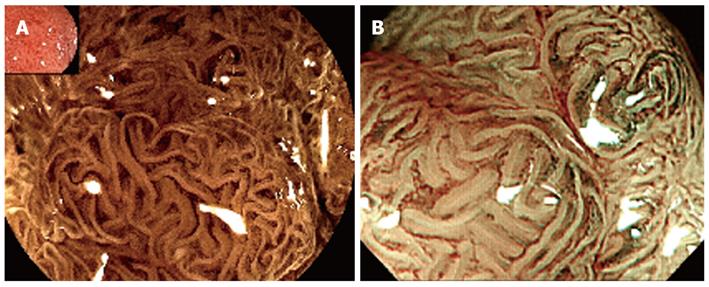
Figure 4 The surface and vascular patterns as seen by flexible spectral imaging color enhancement magnification and narrow band imaging magnification are shown.
A: Flexible spectral imaging color enhancement; B: Narrow band imaging.
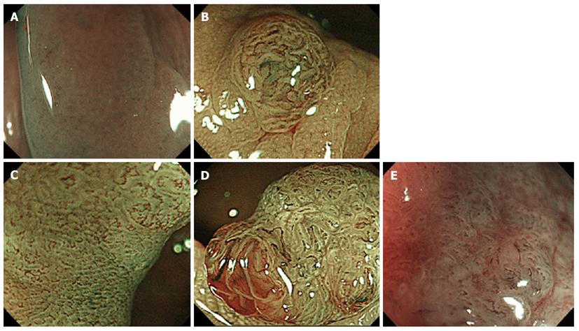
Figure 5 Narrow band imaging classification.
A: Capillary pattern (CP) type I in Sano classification. Type A in Hiroshima classification; B: CP type II in Sano classification. Type B in Hiroshima classification; C: CP type IIIA in Sano classification. Type C1 in Hiroshima classification; D: CP type IIIB in Sano classification. Type C2 in Hiroshima classification; E: CP type IIIB in Sano classification. Type C3 in Hiroshima classification.
Figure 6 The various narrow band imaging classifications and histopathological diagnoses.
Classification of suspect histopathological diagnoses and therapy according to the pattern diagnosed.
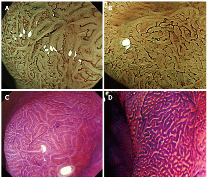
Figure 7 Papillary pattern and tubular pattern in vascular pattern.
A: Papillary type. Papillary pattern is thicker and more winding than the tubular pattern. The surface pattern shows a regular pattern like the IV pit structure according to Hiroshima classification. The pattern was diagnosed type B in Sano classification, type B in Hiroshima classification, Network in Showa classification; B: The pit pattern classification using crystal violet showed IV pit. The histopathological diagnosis of these two patterns shows tubular adenoma; C: Tubular type. Tubular pattern shows a regular honeycomb-like network. The surface pattern shows a regular pattern according to Hiroshima classification. The pattern was diagnosed type B in Sano classification, type B in Hiroshima classification, Network in Showa classification; D: The pit pattern classification using crystal violet showed IIIL pit mainly and IV pit partially. The histopathological diagnosis of these two patterns indicated tubular adenoma.
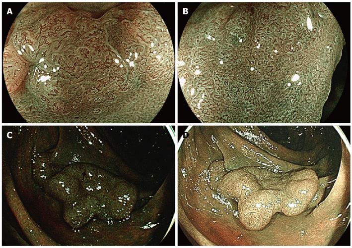
Figure 8 Blue laser imaging.
A: Blue laser imaging (BLI) mode. Clear vascular patterns and the surface pattern can be seen; B: BLI mode. Clear vascular patterns and the surface pattern can be seen; C: BLI mode is slightly dark; D: BLI-bright mode is brighter than BLI mode.
- Citation: Yoshida N, Yagi N, Yanagisawa A, Naito Y. Image-enhanced endoscopy for diagnosis of colorectal tumors in view of endoscopic treatment. World J Gastrointest Endosc 2012; 4(12): 545-555
- URL: https://www.wjgnet.com/1948-5190/full/v4/i12/545.htm
- DOI: https://dx.doi.org/10.4253/wjge.v4.i12.545


