Copyright
©2012 Baishideng Publishing Group Co.
World J Gastrointest Endosc. Oct 16, 2012; 4(10): 462-471
Published online Oct 16, 2012. doi: 10.4253/wjge.v4.i10.462
Published online Oct 16, 2012. doi: 10.4253/wjge.v4.i10.462
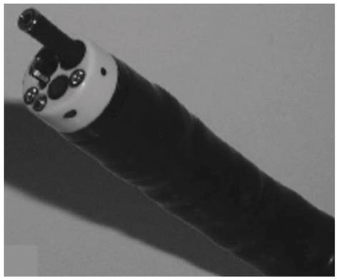
Figure 1 Probe-based endocytoscope being passed through the working channel of a traditional endoscope.
Image from Sasajima et al[4] (used with permission).
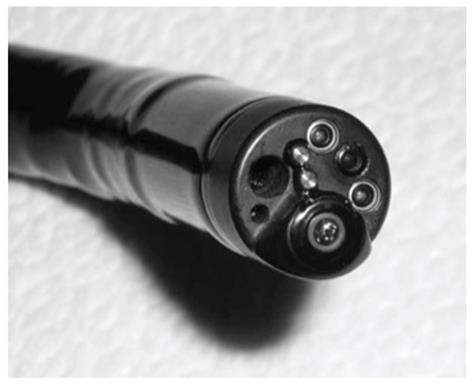
Figure 2 A confocal laser endomicroscope with a 5 mm diameter integrated in the distal end of a traditional colonoscope.
Image from Dekker et al[1] (used with permission).
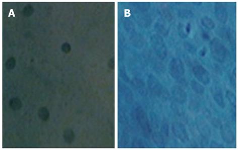
Figure 3 Endocytoscopic images at 1125 × magnification using XEC120U system.
A: Normal squamous cell epithelium of the esophagus with uniform cells; B: Esophageal squamous cell carcinoma. Heterogeneous cells with increased cell density and abnormal nuclei can be seen. Images adapted from Kumagai et al[22] (used with permission).
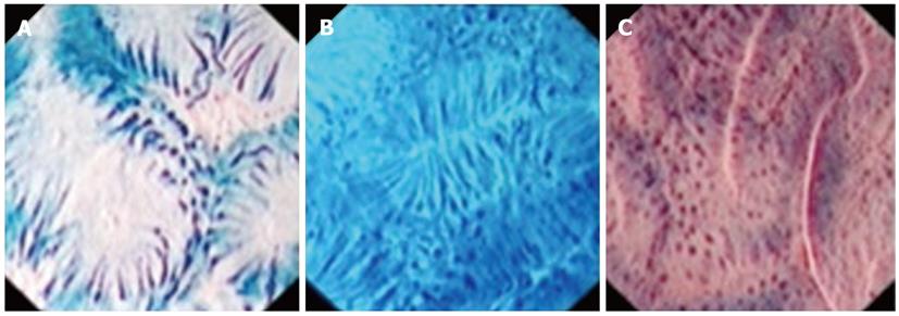
Figure 4 Endocytoscopic images of colorectal neoplasms at 450 × magnification.
A: Low-grade adenoma with elongated nuclei; B: Low-grade adenoma demonstrating elongated nuclei, polygonal crypts, and heterogeneous arrangement; C: High-grade adenoma demonstrating irregular crypts and nuclei that are larger in size and distorted in shape. Images adapted from Rotondano et al[36] (used with permission).
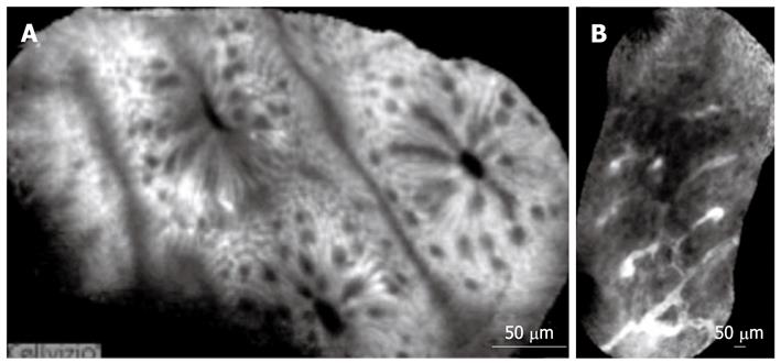
Figure 5 Probe-based confocal laser endomicroscopy.
A: Normal colon; B: Colon adenocarcinoma demonstrating distorted architecture, dilated and irregular vessels, and loss of crypts. Images adapted from De Palma[38] (used with permission).
- Citation: Arya AV, Yan BM. Ultra high magnification endoscopy: Is seeing really believing? World J Gastrointest Endosc 2012; 4(10): 462-471
- URL: https://www.wjgnet.com/1948-5190/full/v4/i10/462.htm
- DOI: https://dx.doi.org/10.4253/wjge.v4.i10.462


