Copyright
©2012 Baishideng Publishing Group Co.
World J Gastrointest Endosc. Oct 16, 2012; 4(10): 438-447
Published online Oct 16, 2012. doi: 10.4253/wjge.v4.i10.438
Published online Oct 16, 2012. doi: 10.4253/wjge.v4.i10.438
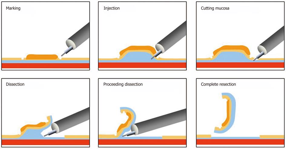
Figure 1 Image of Endoscopic submucosal dissection: Marking is not necessary in a colorectal case because the lesion margins are clear.
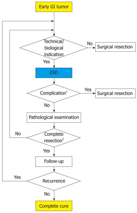
Figure 2 Algorithm for the treatment of early gastrointestinal tumors.
1Perforation or bleeding during endoscopic submucosal dissection which can not be treated endoscopically or delayed perforation; 2See Table 2.
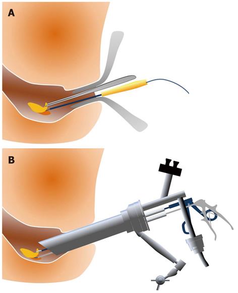
Figure 3 Resection of a rectal tumor.
A: Transanal resection; B: Transanal endoscopic microsurgery.
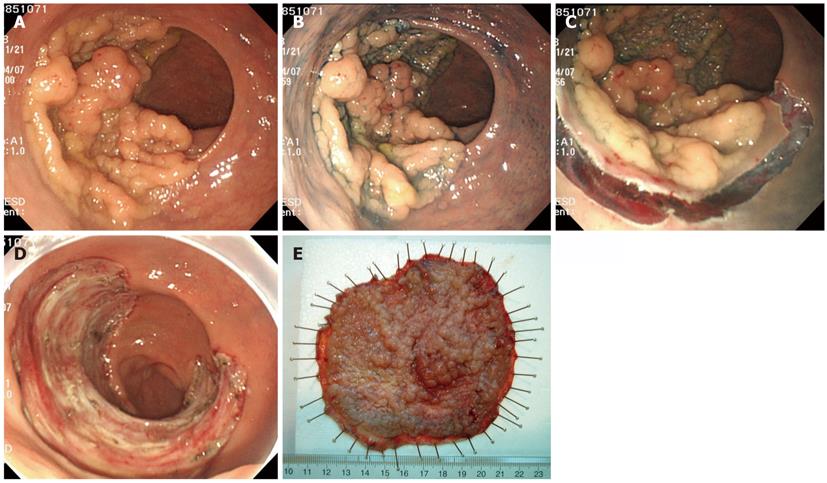
Figure 4 A case of a rectal tumor resected by endoscopic submucosal dissection in which laparotomy was required.
A: A broad-based tumor spreading to over half of the circumference is observed in the rectum; B: Chromoendoscopy with indigo carmine; C: Mucosal incision with the Flush knife; D: Appearance of the mucosa after complete resection by endoscopic submucosal dissection; E: The fixed resected specimen was 115 mm in diameter.
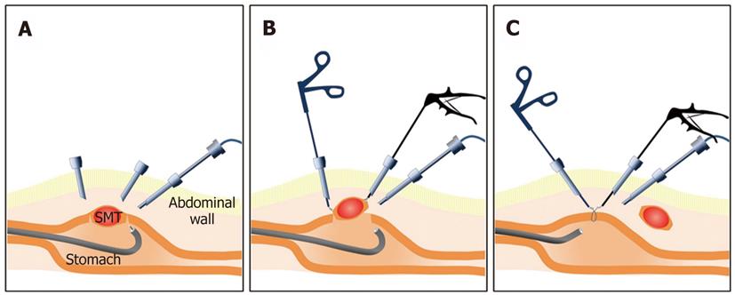
Figure 5 Combination of endoscopic submucosal dissection and laparoscopic surgery.
A: Confirmation of tumor location and mucosal cutting around the tumor using endoscopic submucosal dissection; B: The full thickness of the stomach wall was cut using a laparoscopic instrument, such as Ligasure®; C: The gastric wall was closed using a laparoscopic hand-sewn technique or laparoscopic suturing device, such as End-GIA.
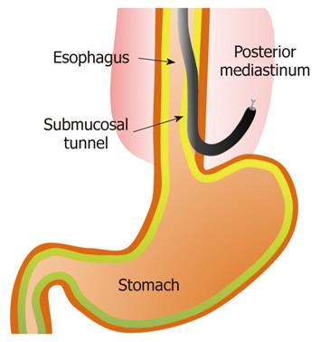
Figure 6 Natural orifice transluminal endoscopic surgery using the endoscopic submucosal dissection technique (in a porcine model).
- Citation: Asano M. Endoscopic submucosal dissection and surgical treatment for gastrointestinal cancer. World J Gastrointest Endosc 2012; 4(10): 438-447
- URL: https://www.wjgnet.com/1948-5190/full/v4/i10/438.htm
- DOI: https://dx.doi.org/10.4253/wjge.v4.i10.438


