Copyright
©2012 Baishideng Publishing Group Co.
World J Gastrointest Endosc. Jan 16, 2012; 4(1): 17-21
Published online Jan 16, 2012. doi: 10.4253/wjge.v4.i1.17
Published online Jan 16, 2012. doi: 10.4253/wjge.v4.i1.17
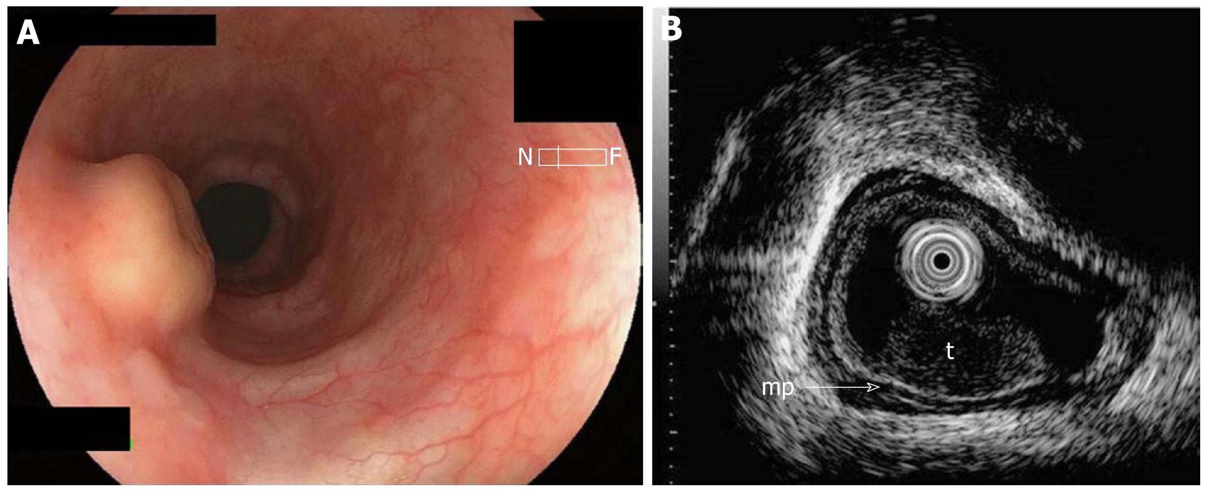
Figure 1 Pretherapeutic examinations of esophageal granular cell tumor.
A: Endoscopic view of the yellowish submucosal tumor with a central depression in the middle esophagus; B: Endoscopic ultrasonography showing a hypoechoic solid tumor (t) in the submucosa; Arrow-mp: Muscularis propria.
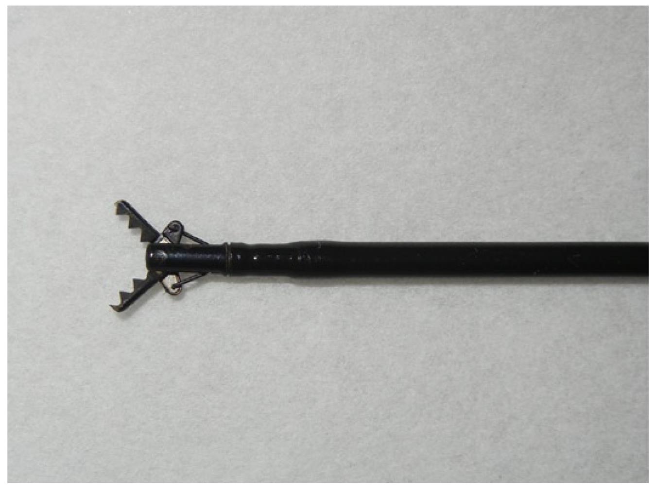
Figure 2 Distal tip of the Clutch Cutter.
The outer side of the forceps is insulated so that electrosurgical current energy is concentrated at the blade to avoid burning the surrounding tissue.
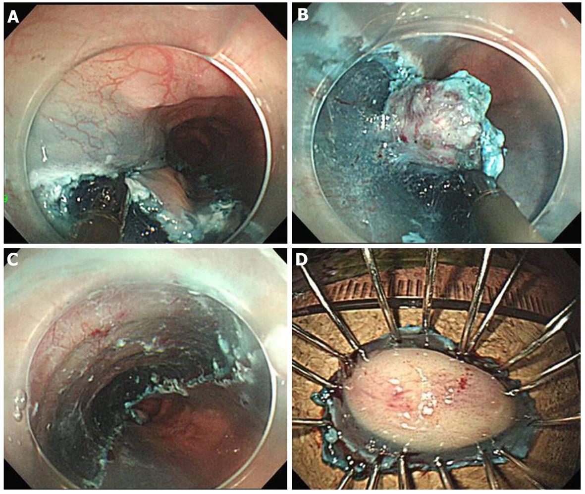
Figure 3 Endoscopic view of the procedure of endoscopic submucosal dissection using the Clutch Cutter.
A: Endoscopic view of partial circumferential incision of the tumor using the Clutch Cutter (CC); B: Endoscopic view of the submucosal exfoliation under the tumor using the CC; C: The lesion is cut completely from the muscle layer; D: The resected specimen showing curative en bloc resection of the lesion.
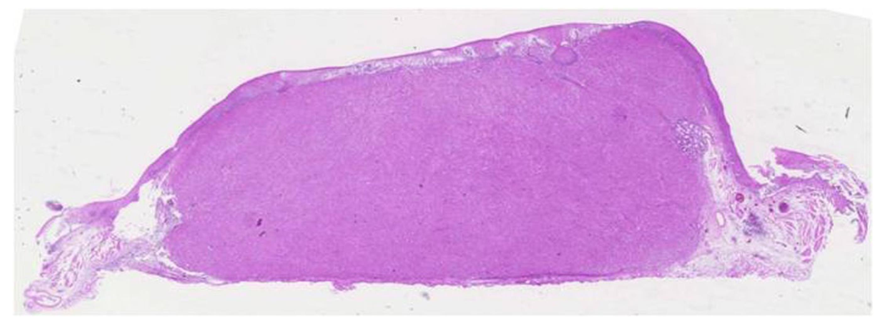
Figure 4 Microscopic appearance of the tumor.
The resected tumor is covered with normal mucosa (hematoxylin and eosin; original magnification, x 2).
- Citation: Komori K, Akahoshi K, Tanaka Y, Motomura Y, Kubokawa M, Itaba S, Hisano T, Osoegawa T, Nakama N, Iwao R, Oya M, Nakamura K. Endoscopic submucosal dissection for esophageal granular cell tumor using the clutch cutter. World J Gastrointest Endosc 2012; 4(1): 17-21
- URL: https://www.wjgnet.com/1948-5190/full/v4/i1/17.htm
- DOI: https://dx.doi.org/10.4253/wjge.v4.i1.17


