Copyright
©2011 Baishideng Publishing Group Co.
World J Gastrointest Endosc. Jul 16, 2011; 3(7): 151-153
Published online Jul 16, 2011. doi: 10.4253/wjge.v3.i7.151
Published online Jul 16, 2011. doi: 10.4253/wjge.v3.i7.151
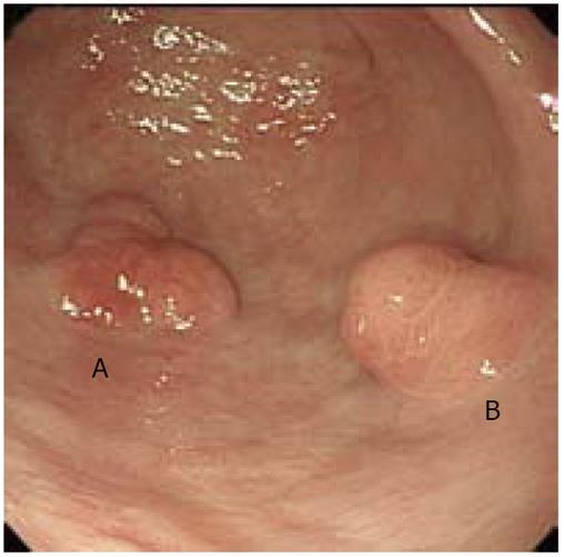
Figure 1 Esophagogastroduodenoscopy findings.
Esophagogastroduod noscopy showed a flat elevated lesion, about 20 mm in size (A), and a polypoid lesion, about 15 mm in size (B), which were detected on the greater curvature of the antrum.
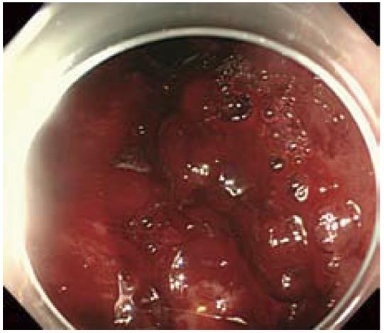
Figure 2 Longitudinal mucosal tears (Mallory-Weiss tear) with active bleeding were detected on the lesser curvature of the gastric corpus.
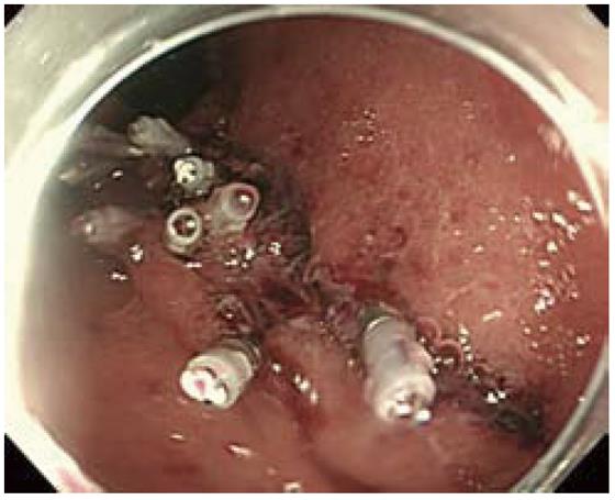
Figure 3 A total of eight hemoclips were applied for hemostasis.
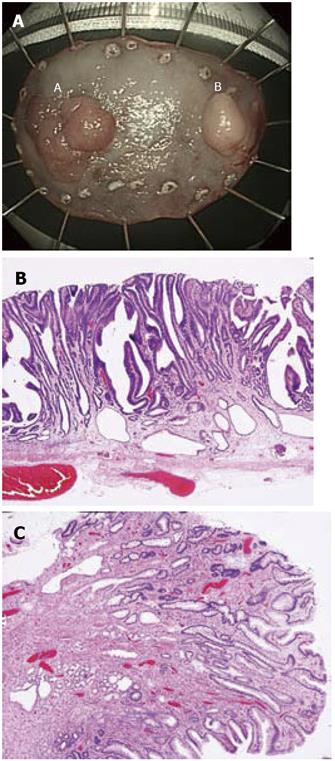
Figure 4 Macroscopic and microscopic findings of resected specimens.
Both lesions were completely removed en bloc (A). Histologically, the first was a papillary carcinoma limited to the mucosal layer and without lymphovascular invasion or involvement of the surgical margins (H&E, × 60) (B), whereas the second lesion was a benign hyperplastic polyp (H&E, × 60) (C).
- Citation: Hongou H, Fu K, Ueyama H, Takahashi T, Takeda T, Miyazaki A, Watanabe S. Mallory-Weiss tear during gastric endoscopic submucosal dissection. World J Gastrointest Endosc 2011; 3(7): 151-153
- URL: https://www.wjgnet.com/1948-5190/full/v3/i7/151.htm
- DOI: https://dx.doi.org/10.4253/wjge.v3.i7.151


