Copyright
©2011 Baishideng Publishing Group Co.
World J Gastrointest Endosc. Jul 16, 2011; 3(7): 133-139
Published online Jul 16, 2011. doi: 10.4253/wjge.v3.i7.133
Published online Jul 16, 2011. doi: 10.4253/wjge.v3.i7.133
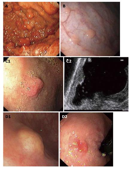
Figure 1 Endoscopic images of early gastrointestinal NETs/carcinoids.
A: Multiple small (< 1 cm), well differentiated (G1) type 2 gastric NETs/carcinoids associated with Zollinger-Ellison-syndrome (ZES) and multiple endocrine neoplasia type 1 (MEN1); B: Multiple small (< 1 cm), well differentiated (G1) type 1 gastric NETs/carcinoids associated with autoimmune chronic atrophic gastritis and pernicious anemia; C: 8 mm measuring NET/carcinoid in the duodenal bulb (C1). Endoscopic ultrasound shows the infiltration of mucosa and submucosa (C2). The duodenal NET/carcinoid exhibits a low echogenic pattern on EUS; D: 10 mm measuring NET/carcinoid of the rectum (D1). 7 mm measuring NET/carcinoid of the rectum (D2). Modified from reference[13-15]. NETs: neuroendocrine tumors; EUS: Endoscopic ultrasound.
- Citation: Scherübl H, Jensen RT, Cadiot G, Stölzel U, Klöppel G. Management of early gastrointestinal neuroendocrine neoplasms. World J Gastrointest Endosc 2011; 3(7): 133-139
- URL: https://www.wjgnet.com/1948-5190/full/v3/i7/133.htm
- DOI: https://dx.doi.org/10.4253/wjge.v3.i7.133


