Copyright
©2011 Baishideng Publishing Group Co.
World J Gastrointest Endosc. Jun 16, 2011; 3(6): 118-123
Published online Jun 16, 2011. doi: 10.4253/wjge.v3.i6.118
Published online Jun 16, 2011. doi: 10.4253/wjge.v3.i6.118
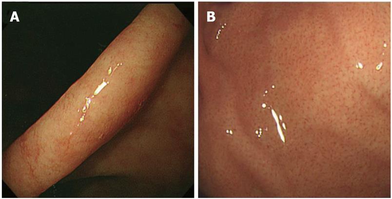
Figure 1 Regular arrangement of collecting venules.
A: Negative, the lower gastric body; B: Positive, the upper gastric body.
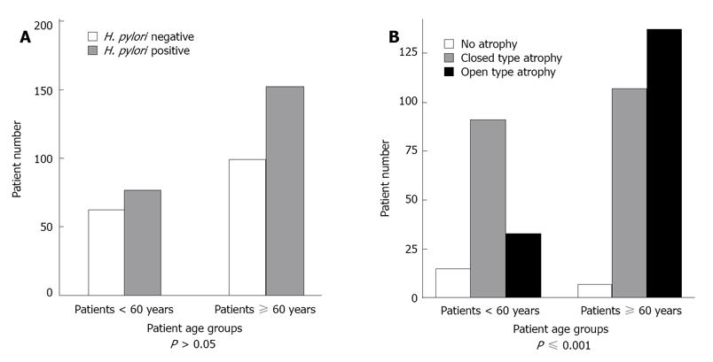
Figure 2 Prevalence of Helicobacter pylori (H.
pylori) infection (A) and distribution of gastric atrophy (B) according to patient age.
- Citation: Alaboudy A, Elbahrawy A, Matsumoto S, Galal GM, Chiba T. Regular arrangement of collecting venules: Does patient age affect its accuracy? World J Gastrointest Endosc 2011; 3(6): 118-123
- URL: https://www.wjgnet.com/1948-5190/full/v3/i6/118.htm
- DOI: https://dx.doi.org/10.4253/wjge.v3.i6.118


