Copyright
©2011 Baishideng Publishing Group Co.
World J Gastrointest Endosc. Feb 16, 2011; 3(2): 46-48
Published online Feb 16, 2011. doi: 10.4253/wjge.v3.i2.46
Published online Feb 16, 2011. doi: 10.4253/wjge.v3.i2.46
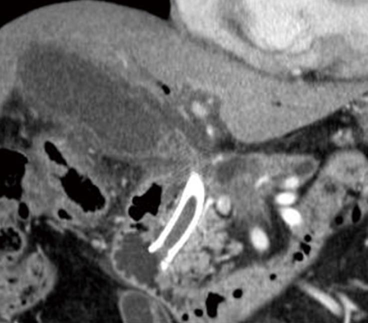
Figure 1 Computed tomography of the abdomen shows swelling of the gallbladder, wall thickening, and the previously placed covered metal stent.
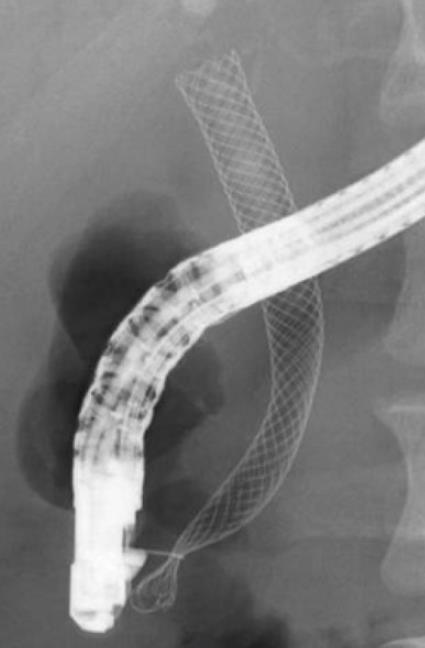
Figure 2 Fluoroscopic image showing the covered self-expandable metal stent grasped with a snare.
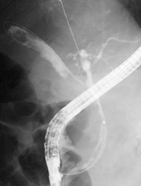
Figure 3 Cholangiogram showing the guidewire being placed in the gallbladder and intrahepatic duct.
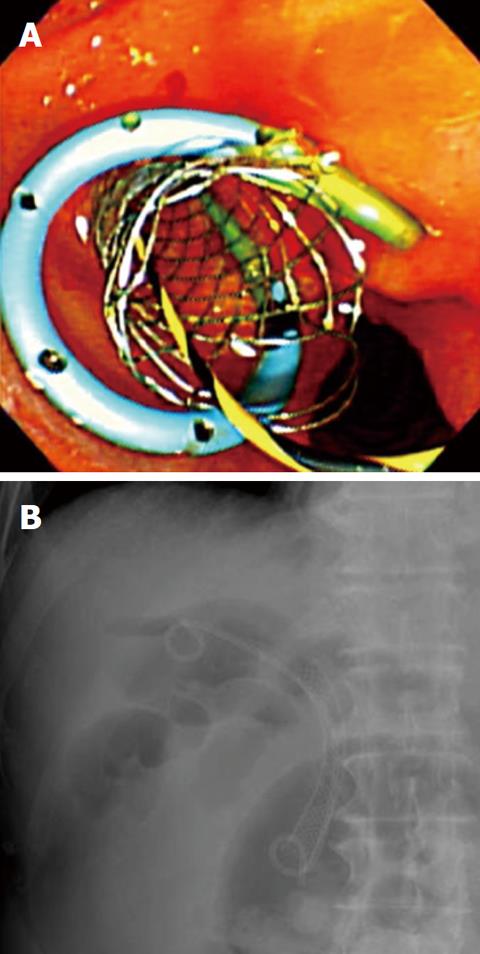
Figure 4 The transpapillary gallbladder stent and covered metal stent.
A: Endoscopic image; B: Fluoroscopic image.
- Citation: Kawakubo K, Isayama H, Sasahira N, Nakai Y, Kogure H, Sasaki T, Hirano K, Tada M, Koike K. Endoscopic transpapillary gallbladder drainage with replacement of a covered self-expandable metal stent. World J Gastrointest Endosc 2011; 3(2): 46-48
- URL: https://www.wjgnet.com/1948-5190/full/v3/i2/46.htm
- DOI: https://dx.doi.org/10.4253/wjge.v3.i2.46


