Copyright
©2011 Baishideng Publishing Group Co.
World J Gastrointest Endosc. Feb 16, 2011; 3(2): 40-45
Published online Feb 16, 2011. doi: 10.4253/wjge.v3.i2.40
Published online Feb 16, 2011. doi: 10.4253/wjge.v3.i2.40
Figure 1 Endoscopic images.
A, B: Video-capsule endoscopy detected a jejunal ulceration without bleeding signs. Subsequently, the patient was transferred to our hospital for further diagnostic work-up; C: We performed double-balloon enteroscopy which confirmed the suspected ulcerous lesion.
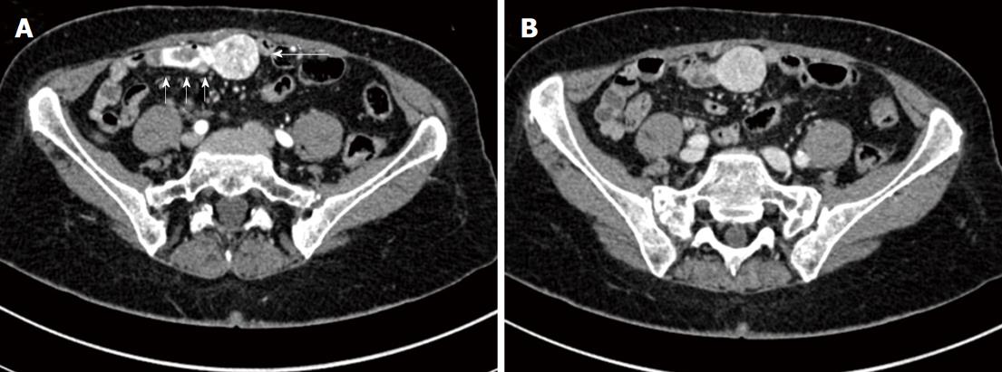
Figure 2 Biphasic MDCT in the arterial and venous phase.
A: Smoothly outlined abdo-minal mass (transverse diameter of about 2.8 cm x 3 cm, long arrow) showing considerable arterial contrast enhancement and intraluminal contrast medium extravasation (shot arrows); B: Washout in the venous phase.
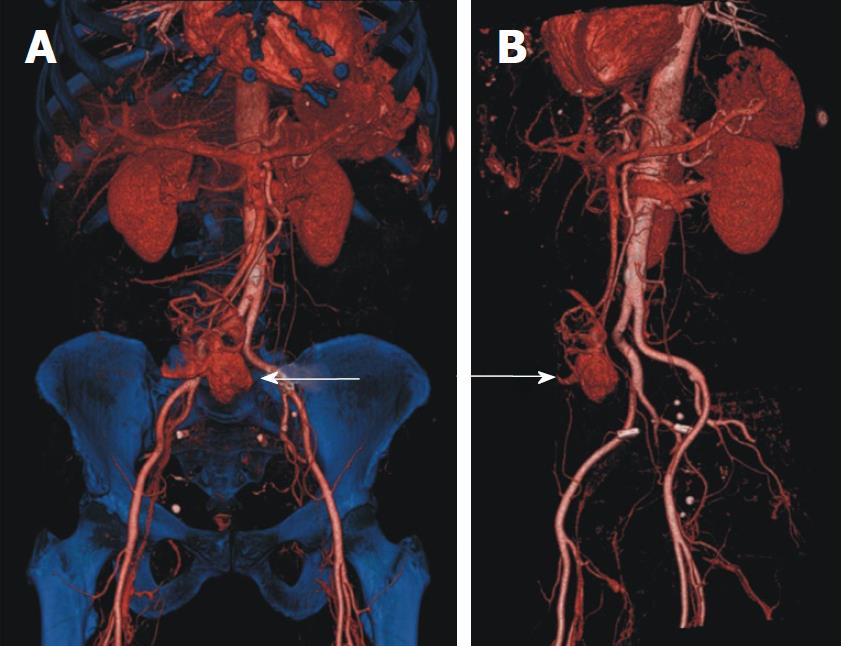
Figure 3 3D volume rendered reconstructions in the arterial phase (A, B).
Note that the tumour (arrows) is supplied by vessels from the superior mesenteric and iliac arterial territories.
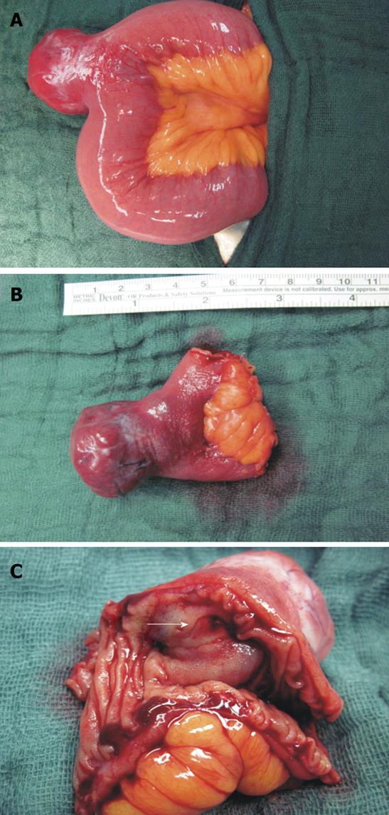
Figure 4 Surgical specimens.
A: Tumor of 3.6 cm × 3.2 cm × 2.8 cm arising from the jejunum (240 cm behind the plica duodenalis superior/ Treitz’s ligament); B:removal of the tumor via a partial jejunal resection; C: sliced preparation of the jejunum with a view of the GIST related ulcer (arrow).
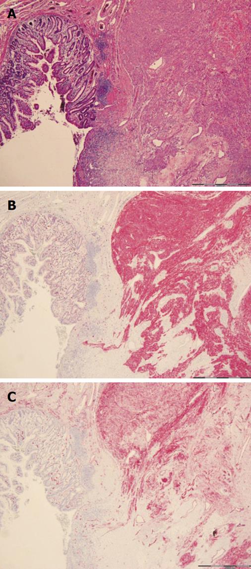
Figure 5 Histological assessment of surgical specimen revealed an ulcerated spindle-celled gastric stromal tumor with well marked margin and a positive staining for CD117 and CD34.
A: HE-; B: CD117-; C: CD34-staining.
- Citation: Reuter S, Bettenworth D, Mees ST, Neumann J, Beyna T, Domschke W, Wessling J, Ullerich H. A typical presentation of a rare cause of obscure gastrointestinal bleeding. World J Gastrointest Endosc 2011; 3(2): 40-45
- URL: https://www.wjgnet.com/1948-5190/full/v3/i2/40.htm
- DOI: https://dx.doi.org/10.4253/wjge.v3.i2.40


