Copyright
©2010 Baishideng Publishing Group Co.
World J Gastrointest Endosc. Sep 16, 2010; 2(9): 321-324
Published online Sep 16, 2010. doi: 10.4253/wjge.v2.i9.321
Published online Sep 16, 2010. doi: 10.4253/wjge.v2.i9.321
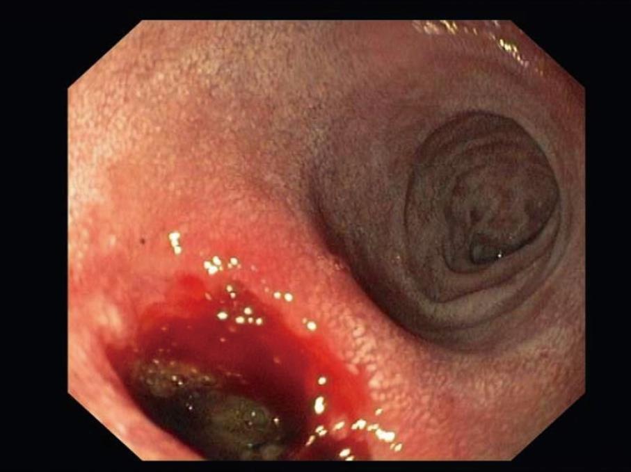
Figure 1 Upper gastrointestinal endoscopy showing a deep ulcer of the duodenal bulb.
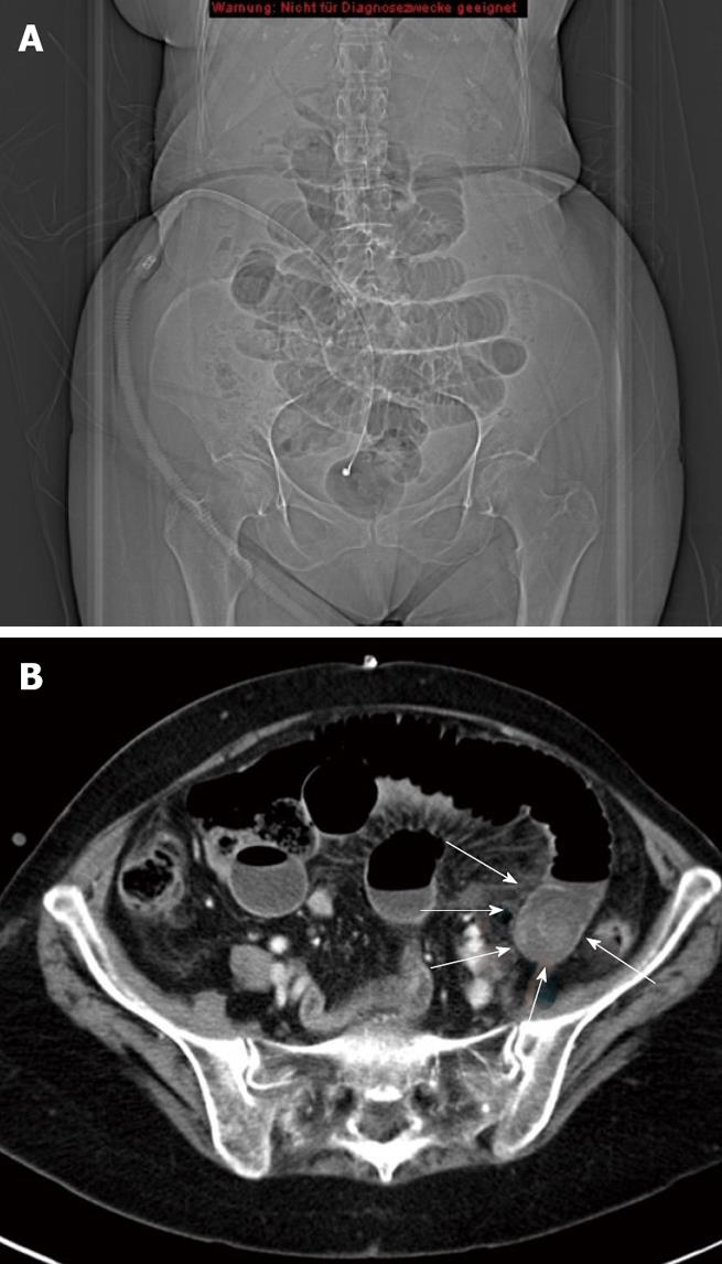
Figure 2 Abdominal computed tomography-scan.
A: The typical image of mechanical small bowel obstruction with distended small bowel loops reaching to the mid ileum as well as pneumobilia of the central biliary tract; B: A calcified mass of 5 cm in diameter located in the distal small bowel.
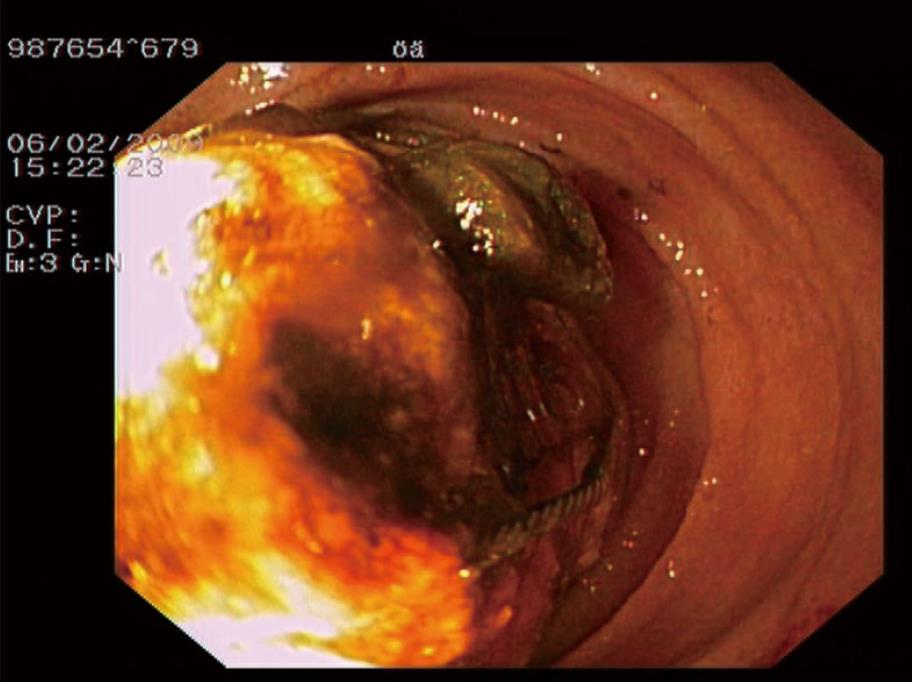
Figure 3 Single-balloon enteroscopy via the oral route revealing a calculus, about 450 cm distant from the pylorus, completely occluding the intestinal lumen.
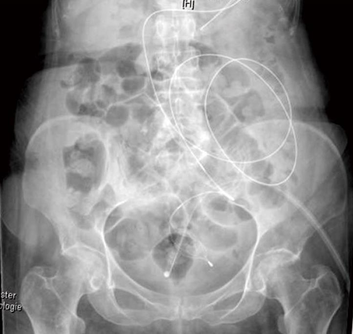
Figure 4 Plain X-ray showing an ileal calculus endoscopically captured with a dormia basket guided by single-balloon enteroscopy.
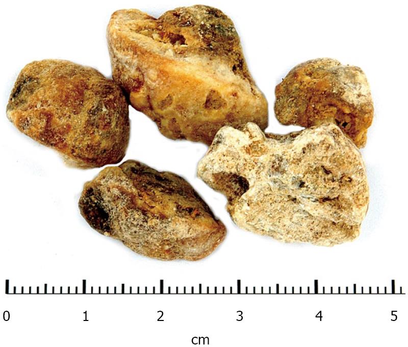
Figure 5 Removed gallstone fragments.
- Citation: Heinzow HS, Meister T, Wessling J, Domschke W, Ullerich H. Ileal gallstone obstruction: Single-balloon enteroscopic removal. World J Gastrointest Endosc 2010; 2(9): 321-324
- URL: https://www.wjgnet.com/1948-5190/full/v2/i9/321.htm
- DOI: https://dx.doi.org/10.4253/wjge.v2.i9.321


