Copyright
©2010 Baishideng Publishing Group Co.
World J Gastrointest Endosc. Sep 16, 2010; 2(9): 314-317
Published online Sep 16, 2010. doi: 10.4253/wjge.v2.i9.314
Published online Sep 16, 2010. doi: 10.4253/wjge.v2.i9.314
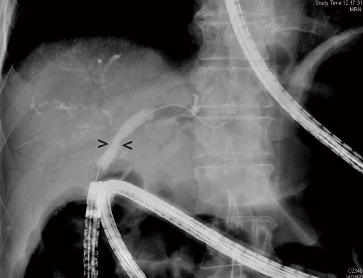
Figure 1 Radiologic view of a balloon dilation of a stenotic hepaticojejunostomy.
The arrows indicate the stenosis.
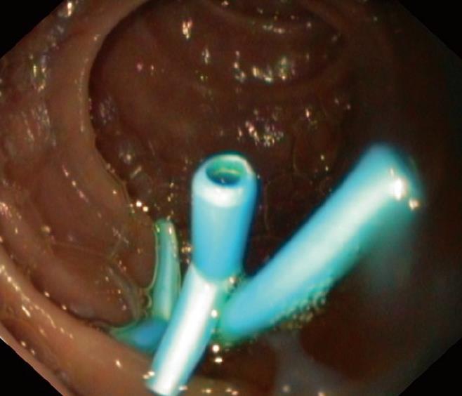
Figure 2 Three 7 Fr stents were placed through the enteroscope in the enterobiliary anastomosis after balloon dilation of the postoperative stenosis.
Bile is spontaneously evacuated through and between the stents.
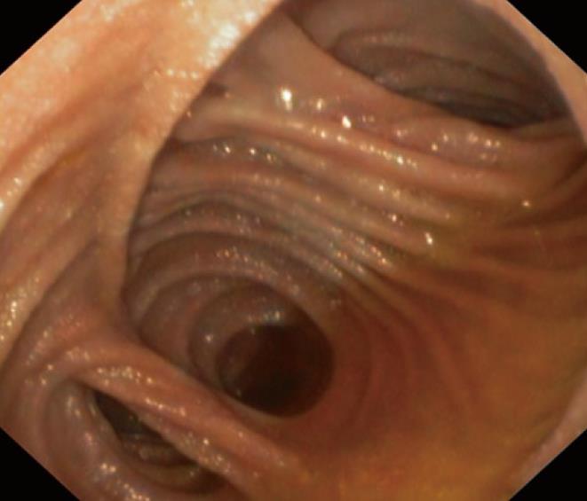
Figure 3 Endoscopic view of a side-to-side Roux-en-Y entero-enteric anastomosis, showing 3 different directions of jejunal limbs.
The upper right directs towards the afferent Roux-en-Y limb. This limb often contains bile and presents antiperistaltic contractions.
- Citation: Moreels TG, Pelckmans PA. Comparison between double-balloon and single-balloon enteroscopy in therapeutic ERC after Roux-en-Y entero-enteric anastomosis. World J Gastrointest Endosc 2010; 2(9): 314-317
- URL: https://www.wjgnet.com/1948-5190/full/v2/i9/314.htm
- DOI: https://dx.doi.org/10.4253/wjge.v2.i9.314


