Copyright
©2010 Baishideng.
World J Gastrointest Endosc. Aug 16, 2010; 2(8): 293-297
Published online Aug 16, 2010. doi: 10.4253/wjge.v2.i8.293
Published online Aug 16, 2010. doi: 10.4253/wjge.v2.i8.293
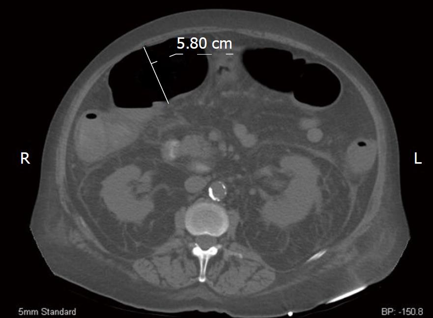
Figure 1 Computerized tomography axial image of our patient.
We measured the patient’s transverse colon at 5.80 cm. Additionally, perinephric fluid is markedly evident bilaterally.
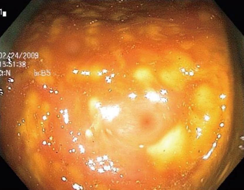
Figure 2 Colonoscopic image taken of the descending colon consistent with pseudomembranous colitis.
-
Citation: Sayedy L, Kothari D, Richards RJ. Toxic megacolon associated
Clostridium difficile colitis. World J Gastrointest Endosc 2010; 2(8): 293-297 - URL: https://www.wjgnet.com/1948-5190/full/v2/i8/293.htm
- DOI: https://dx.doi.org/10.4253/wjge.v2.i8.293


