Copyright
©2010 Baishideng.
World J Gastrointest Endosc. Jul 16, 2010; 2(7): 244-251
Published online Jul 16, 2010. doi: 10.4253/wjge.v2.i7.244
Published online Jul 16, 2010. doi: 10.4253/wjge.v2.i7.244
Figure 1 Pill cam colon capsule.
A: a normal colon (cecum and ileocecal valve); B: A small colonic diverticulum in the colon transversum; C: The rectum with internal haemorrhoids (equivalent to a retroflex view with the standard colonoscope).
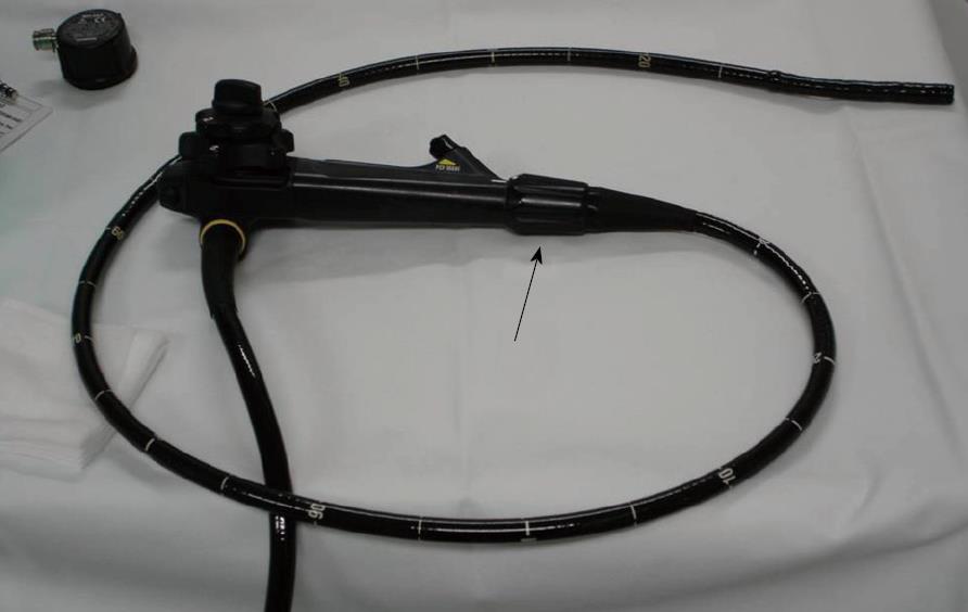
Figure 2 Variable stiffness colonoscope with the control ring to adjust stiffness (arrow).
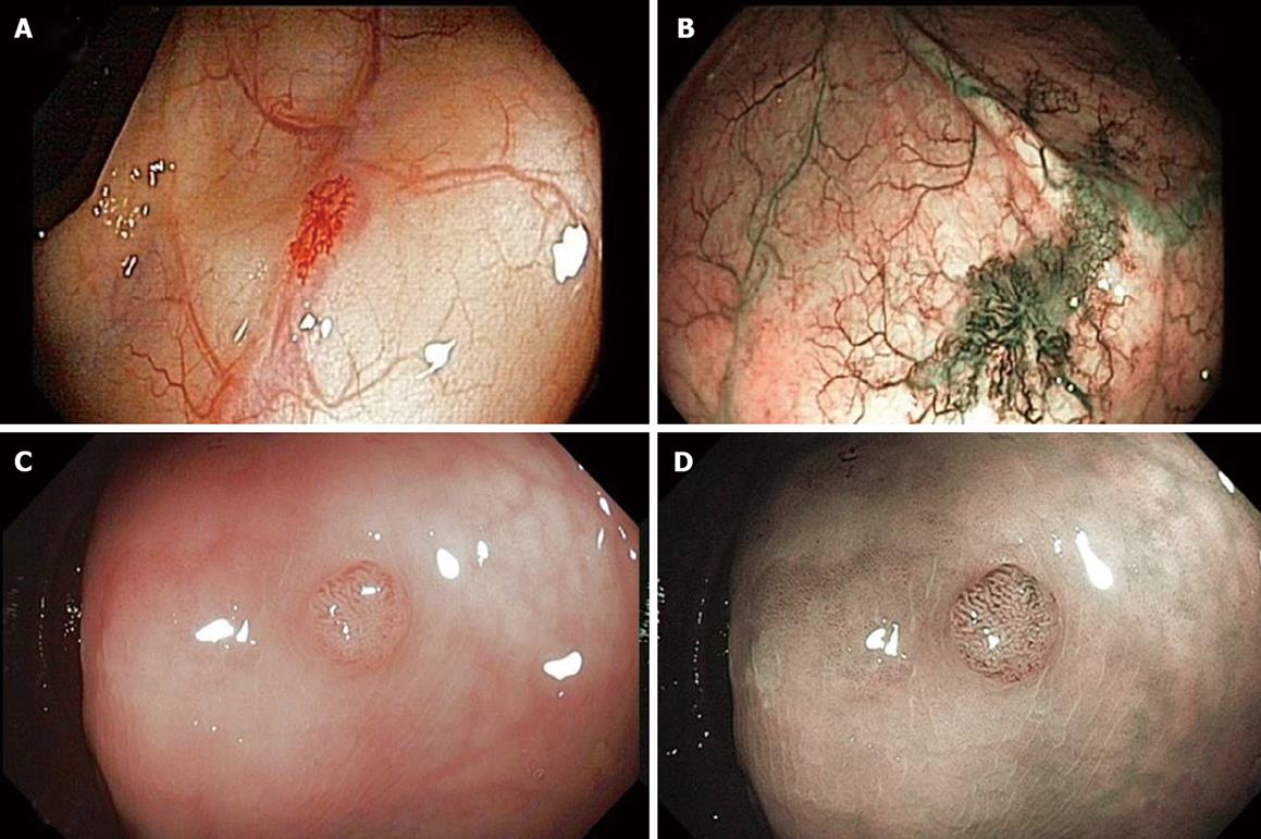
Figure 3 Visualization of colonic lesions.
A: Angiodysplasias under standard view; B: Angiodysplasias under improved visualization with narrow–band Imaging (NBI); C: A colonic polyp displayed with standard colonoscopy; D: The same polyp with NBI.
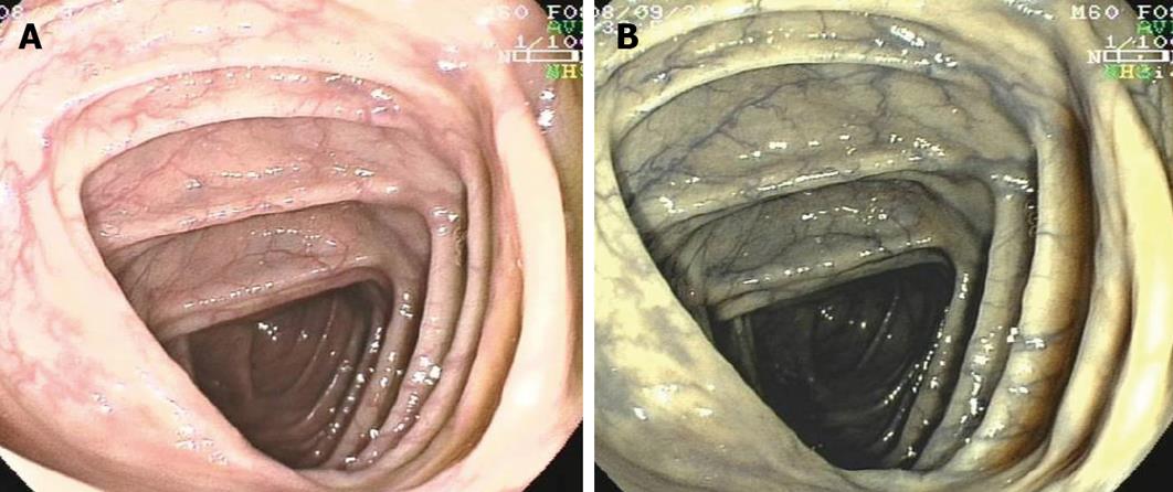
Figure 4 Visualization of the colon transversum.
A: Under standard view; B: With Fujinon intelligent chromoendoscopy (FICE). Note the improved visualization of the capillary pattern of the mucosa with FICE.
- Citation: Gaglia A, Papanikolaou IS, Veltzke-Schlieker W. New endoscopy devices to improve population adherence to colorectal cancer prevention programs. World J Gastrointest Endosc 2010; 2(7): 244-251
- URL: https://www.wjgnet.com/1948-5190/full/v2/i7/244.htm
- DOI: https://dx.doi.org/10.4253/wjge.v2.i7.244


