Copyright
©2010 Baishideng.
World J Gastrointest Endosc. Mar 16, 2010; 2(3): 90-96
Published online Mar 16, 2010. doi: 10.4253/wjge.v2.i3.90
Published online Mar 16, 2010. doi: 10.4253/wjge.v2.i3.90
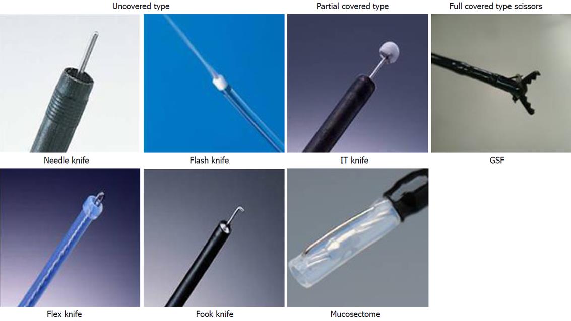
Figure 1 Several endoscopic cutting devices have been developed in Japan.
They are divided into 3 broad categories as above: Uncovered type knife, partial-covered type knife, and full-covered type scissors.
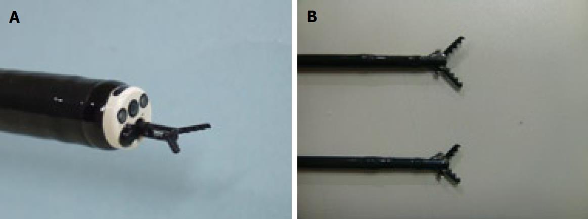
Figure 2 Grasping type scissor forceps (GSF).
A: Distal tip of the long type GSF. The outer side of the forceps is insulated so that electrosurgical current energy is concentrated at the blade to avoid burning to the surrounding tissue (closed discharge). B: Long type (upper side image: 6mm) and short type (lower side image: 4 mm) GSF.
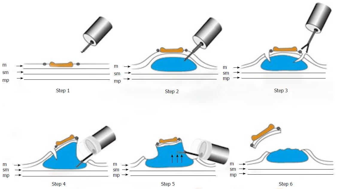
Figure 3 Schematic shows ESD using GSF.
Step 1: Marking dots are made on the circumference of the lesion to outline the incision line; Step 2: A concentrated glycerin solution mixed with a small volume of epinephrine and indigo carmine dye is injected into the submucosal layer around the target lesion to lift the entire lesion; Step 3: The lesion is separated from the surrounding normal mucosa by complete incision around the lesion using the GSF; Step 4: A piece of submucosal tissue is grasped by GSF; Step5: A grasped tissue is lifted up and cut with the GSF using electrosurgical current to effect submucosal exfoliation; Step 6: The lesion is resected in one piece; m: Mucosa; sm: Submucosa; mp: Muscularis propria.
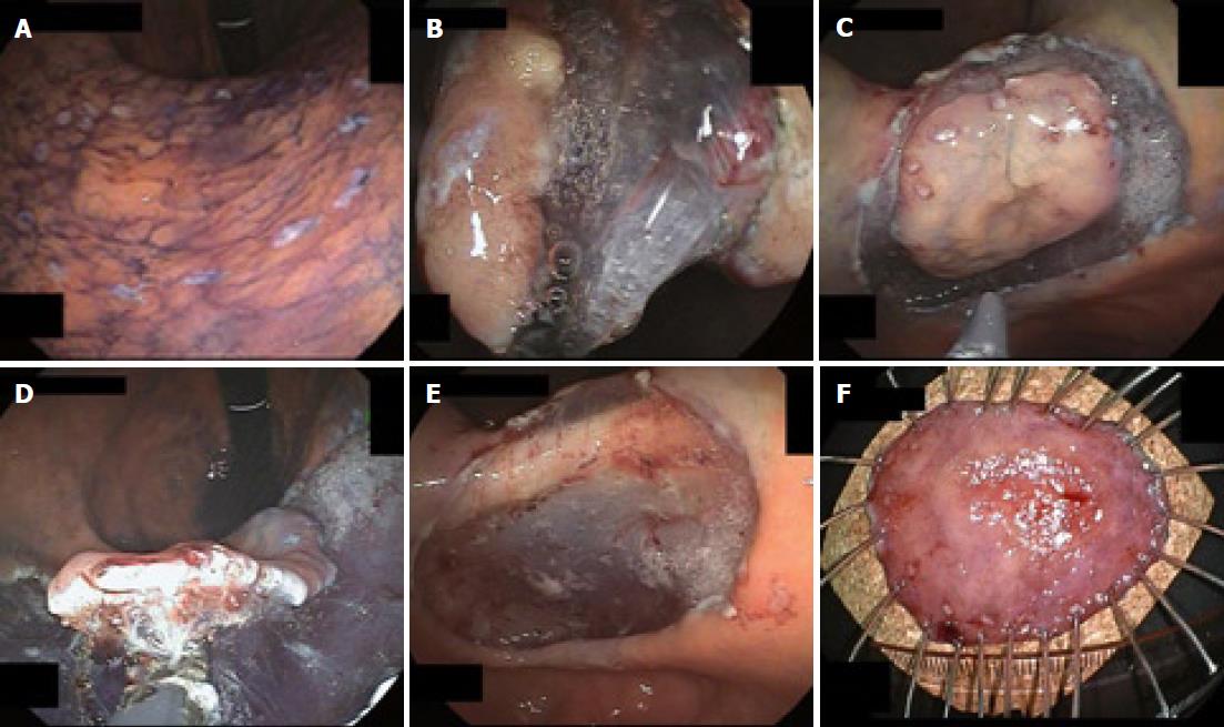
Figure 4 Endoscopic view of the procedure of ESD using GSF.
A: Marks are made at several points along the outline of the lesion with a coagulation current; B: The mucosa is incised outside the marker dots to separate the lesion from the surrounding non-neoplastic mucosa using GSF; C: Completion of the GSF cutting around the lesion with a safe lateral margin; D: The submucosal connective tissue beneath the lesion is grasped and lifted up and excised using GSF from the underlying muscle layer; E: The lesion is cut completely from the muscle layer; F: The resected specimen showing en bloc resection of the lesion.
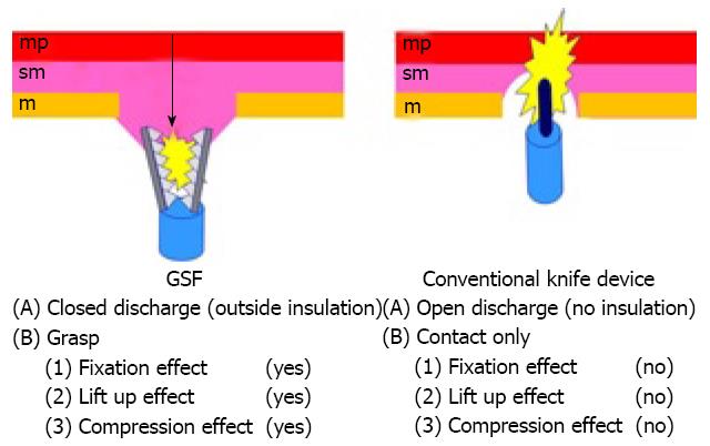
Figure 5 Mechanical differences between GSF and conventional knife devices.
sm: Submucosa; mp: Muscularis propria.
- Citation: Akahoshi K, Akahane H. A new breakthrough: ESD using a newly developed grasping type scissor forceps for early gastrointestinal tract neoplasms. World J Gastrointest Endosc 2010; 2(3): 90-96
- URL: https://www.wjgnet.com/1948-5190/full/v2/i3/90.htm
- DOI: https://dx.doi.org/10.4253/wjge.v2.i3.90


