Copyright
©2010 Baishideng Publishing Group Co.
World J Gastrointest Endosc. Nov 16, 2010; 2(11): 379-380
Published online Nov 16, 2010. doi: 10.4253/wjge.v2.i11.379
Published online Nov 16, 2010. doi: 10.4253/wjge.v2.i11.379
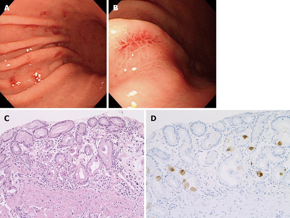
Figure 1 Endoscopic and histopathological pictures of cytomegalovirus gastritis.
A: Note numerous patchy erythemas in the gastric body; B: Closer observation showing the slightly depressed erythema; C: Histopathological examination of the erythema showing large epithelial cells with characteristic “owl’s eye” eosinophilic intranuclear inclusion body surrounded by a clear halo (H&E, × 200); D: Note positive immunostaining for cytomegalovirus antigens × 200).
- Citation: Hokama A, Taira K, Yamamoto YI, Kinjo N, Kinjo F, Takahashi K, Fujita J. Cytomegalovirus gastritis. World J Gastrointest Endosc 2010; 2(11): 379-380
- URL: https://www.wjgnet.com/1948-5190/full/v2/i11/379.htm
- DOI: https://dx.doi.org/10.4253/wjge.v2.i11.379


