Copyright
©The Author(s) 2025.
World J Gastrointest Endosc. Jun 16, 2025; 17(6): 104539
Published online Jun 16, 2025. doi: 10.4253/wjge.v17.i6.104539
Published online Jun 16, 2025. doi: 10.4253/wjge.v17.i6.104539
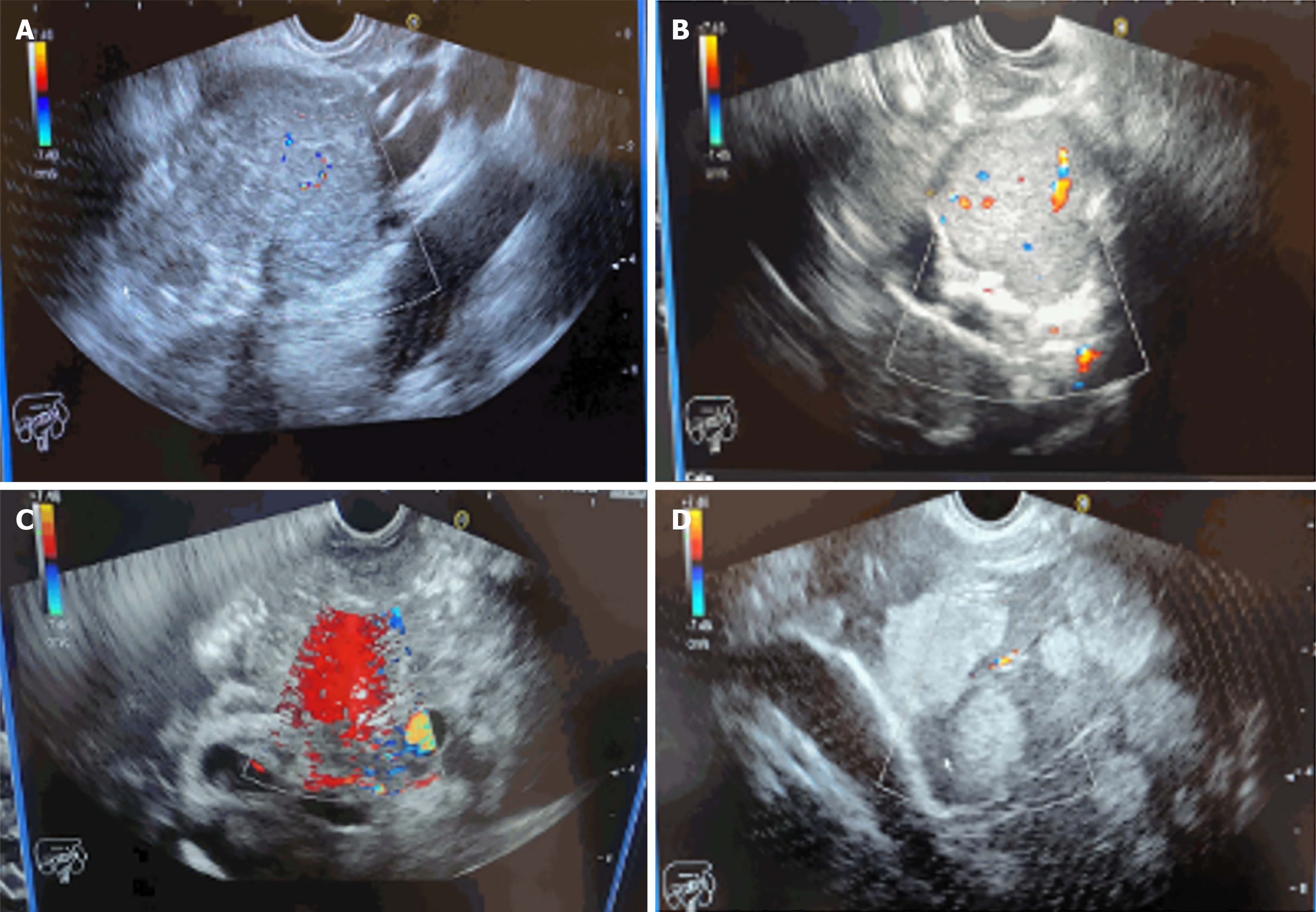
Figure 1 Endoscopic ultrasound demonstration of neuroendocrine tumors at various gastrointestinal sites in different patients.
A: Pancreatic mass located in the head region; B: Well-defined, vascularized porta hepatis mass; C: Hypoechoic mediastinal mass; D: Multiple hyperechoic enhancing liver lesions.
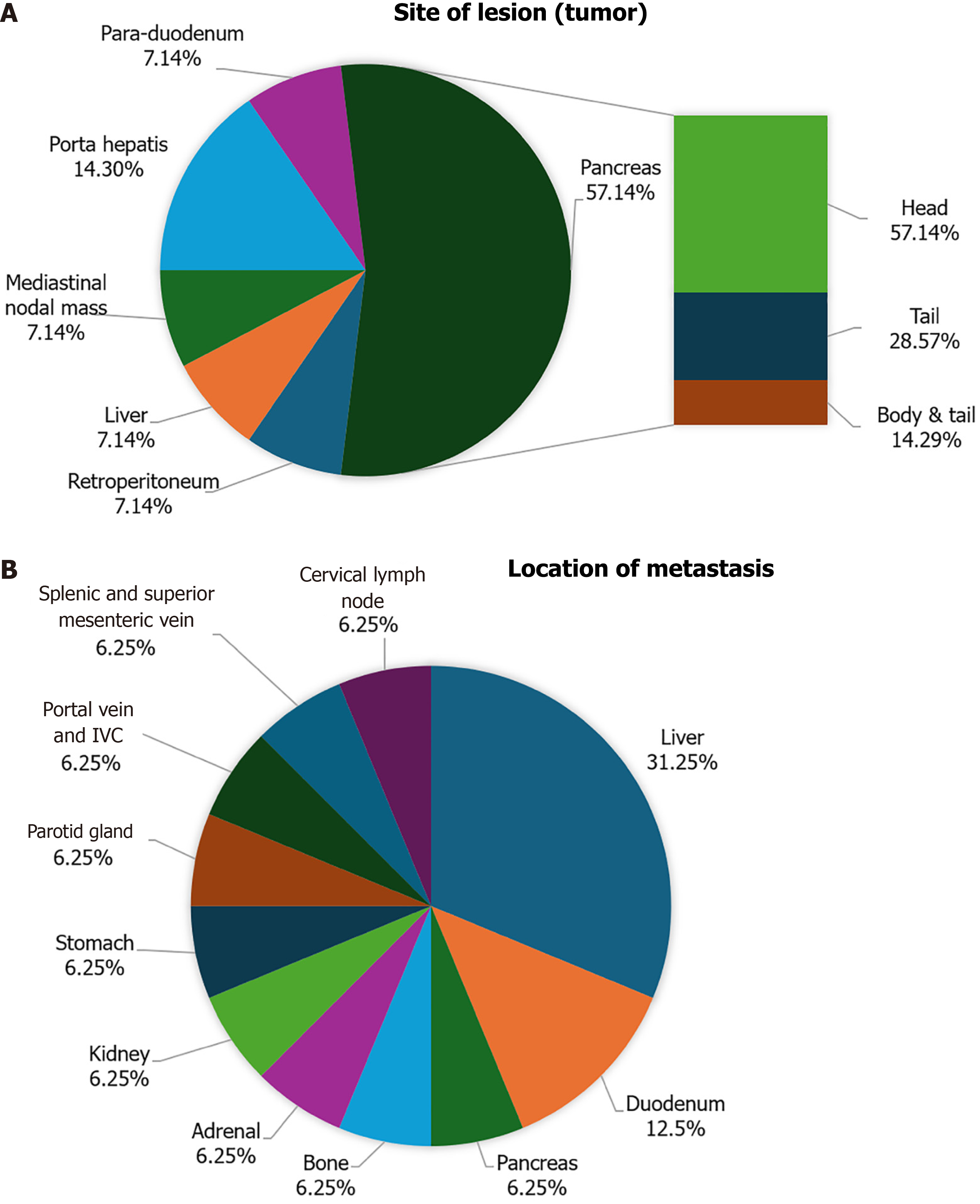
Figure 2 The distribution of gastroenteropancreatic neuroendocrine tumors based on the primary lesion site and metastatic sites among patients with gastroenteropancreatic-neuroendocrine tumors.
A: The distribution of gastroenteropancreatic (GEP) neuroendocrine tumors (NETs) based on the primary lesion site. The majority of tumors were located in the pancreas, with further sub-classification showing that most pancreatic lesions were found in the head region. These findings are based on imaging and histologic assessments; B: The distribution of metastatic sites among patients with GEP-NETs. The liver was the most common site of metastasis, accounting for 32% of all metastatic cases.
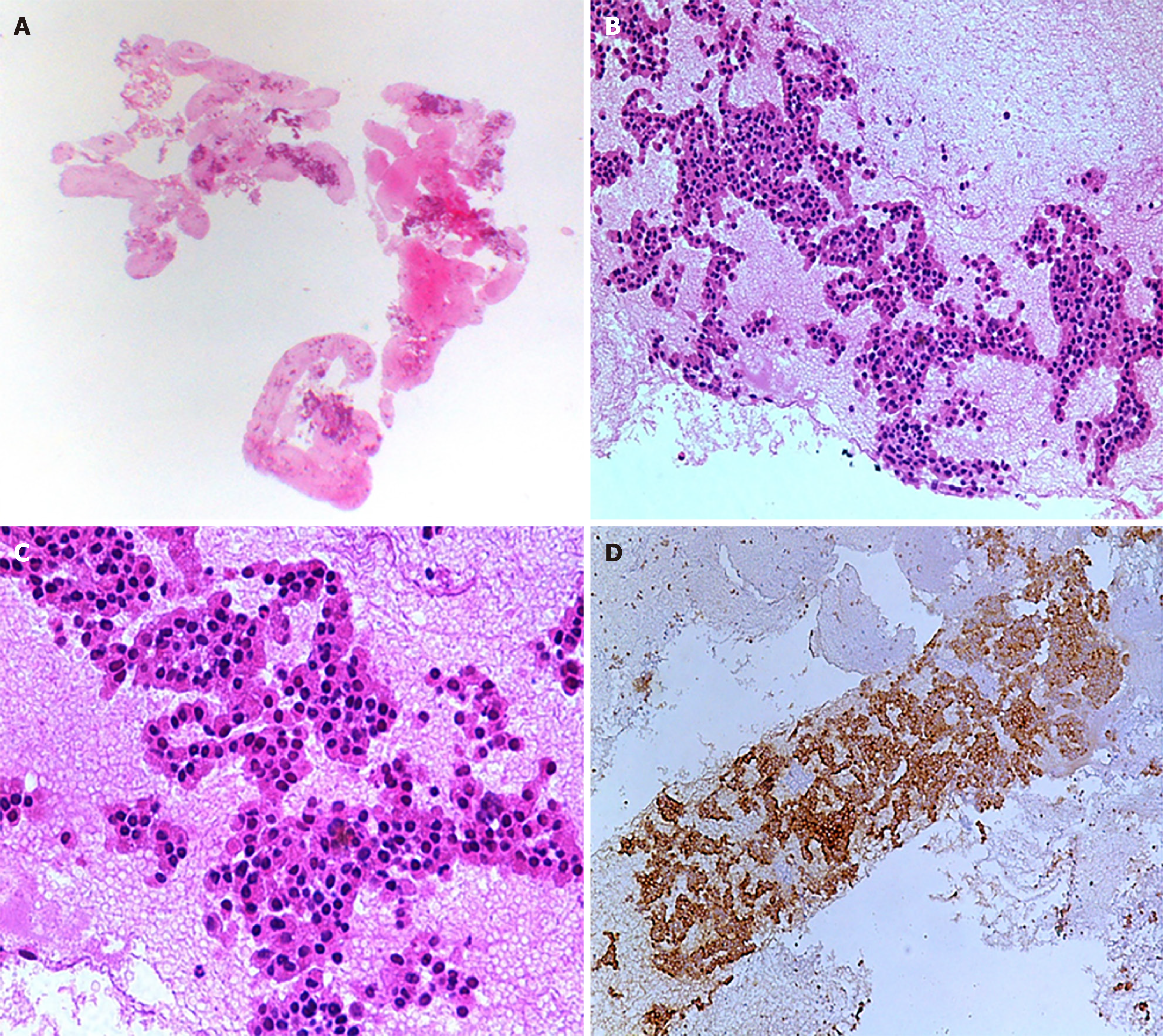
Figure 3 Photomicrographs of a neuroendocrine tumor in the pancreatic uncinate mass.
A and B: Low- and intermediate-power views of the endoscopic ultrasonography-guided biopsy sample, revealing a tumor composed of epithelial cells arranged in nests and trabeculae. C: Tumor cells with monotonous round nuclei, hyperchromasia, and no significant atypia or mitoses; D: These morphological findings, along with immunohistochemical expression of the neuroendocrine marker synaptophysin, epithelial marker cytokeratin, and a Ki-67 proliferative index of < 2%, are consistent with a well-differentiated neuroendocrine tumor, World Health Organization grade I (H&E 4 ×, 10 ×, 20 × & immunohistochemistry).
- Citation: Ayesha S, Karim MM, Shahid AH, Rehman AU, Uddin Z, Abid S. Diagnostic role of endoscopic ultrasonography in defining the clinical features and histopathological spectrum of gastroenteropancreatic neuroendocrine tumors. World J Gastrointest Endosc 2025; 17(6): 104539
- URL: https://www.wjgnet.com/1948-5190/full/v17/i6/104539.htm
- DOI: https://dx.doi.org/10.4253/wjge.v17.i6.104539


