Copyright
©The Author(s) 2025.
World J Gastrointest Endosc. Feb 16, 2025; 17(2): 101135
Published online Feb 16, 2025. doi: 10.4253/wjge.v17.i2.101135
Published online Feb 16, 2025. doi: 10.4253/wjge.v17.i2.101135
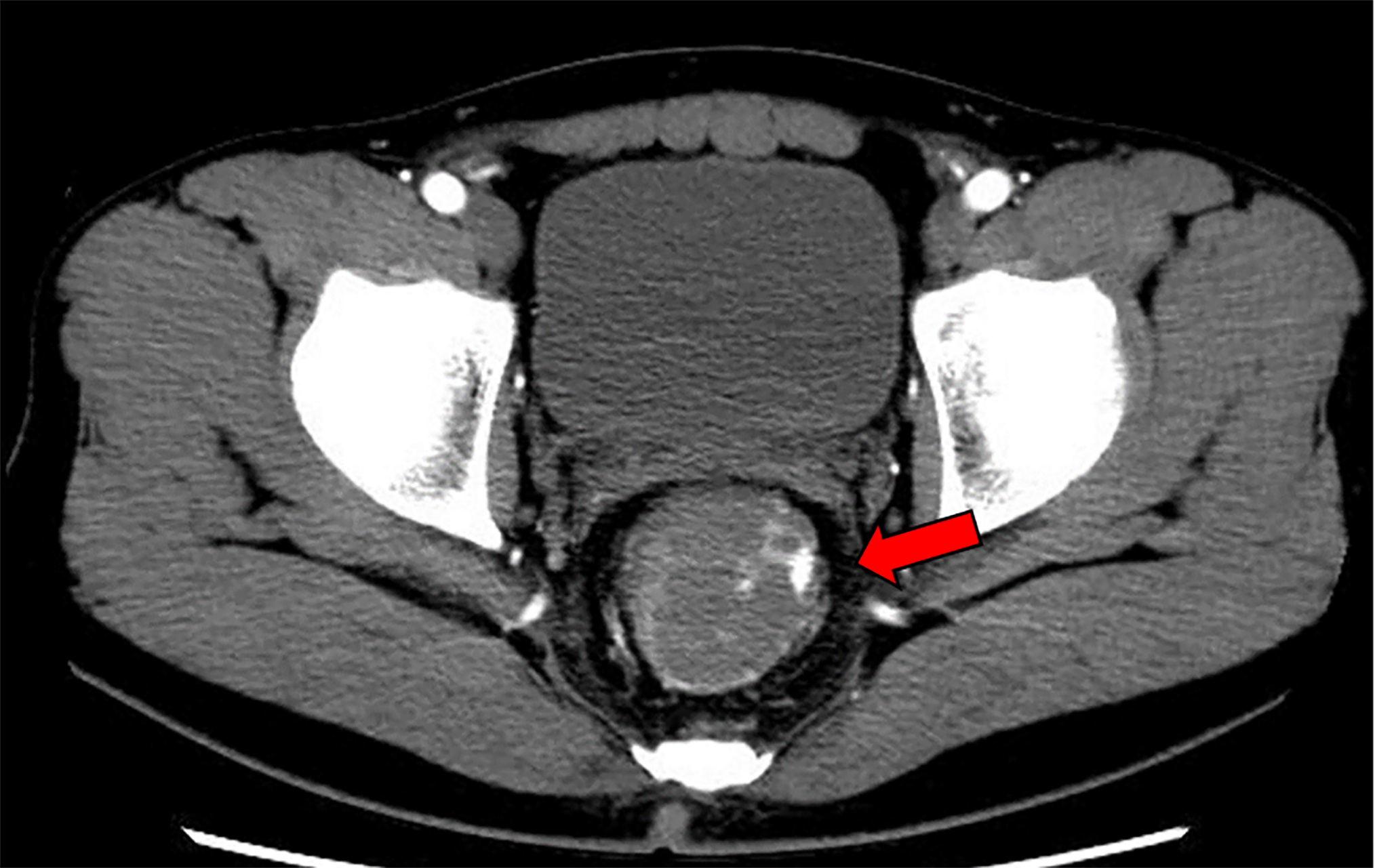
Figure 1 Contrast-enhanced abdominal computed tomography scan.
Contrast-enhanced abdominal computed tomography scan shows extravascular leakage of the contrast medium from the left wall of the lower rectum (red arrow).
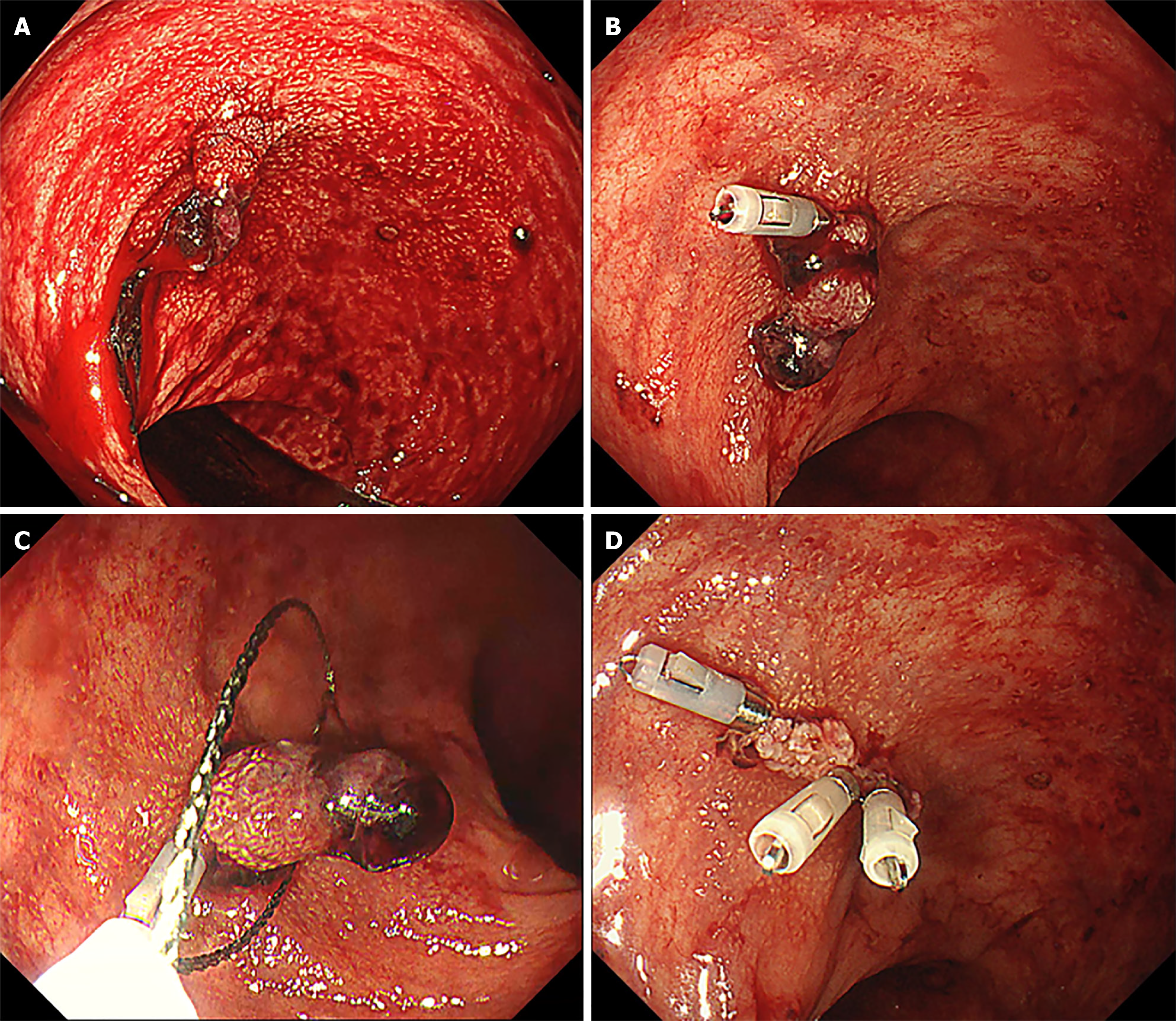
Figure 2 Endoscopic hemostasis and polypectomy.
A: Pulsatile bleeding is observed from the stem of the polyp; B: Endoscopic hemostasis is performed by clipping the stem; C: Polypectomy is then performed above the clip to examine the tissue; D: The clips are added to completely stop the bleeding.
Figure 3 Histopathological evaluation.
A: Hematoxylin and eosin staining showing large blood vessels in the stalk (red box) and some dilated glandular ducts on the cyst (yellow arrowheads); B: Desmin staining showing intricate lamina muscularis mucosae. There are no signs of malignancy, such as tearing or disruption of the lamina.
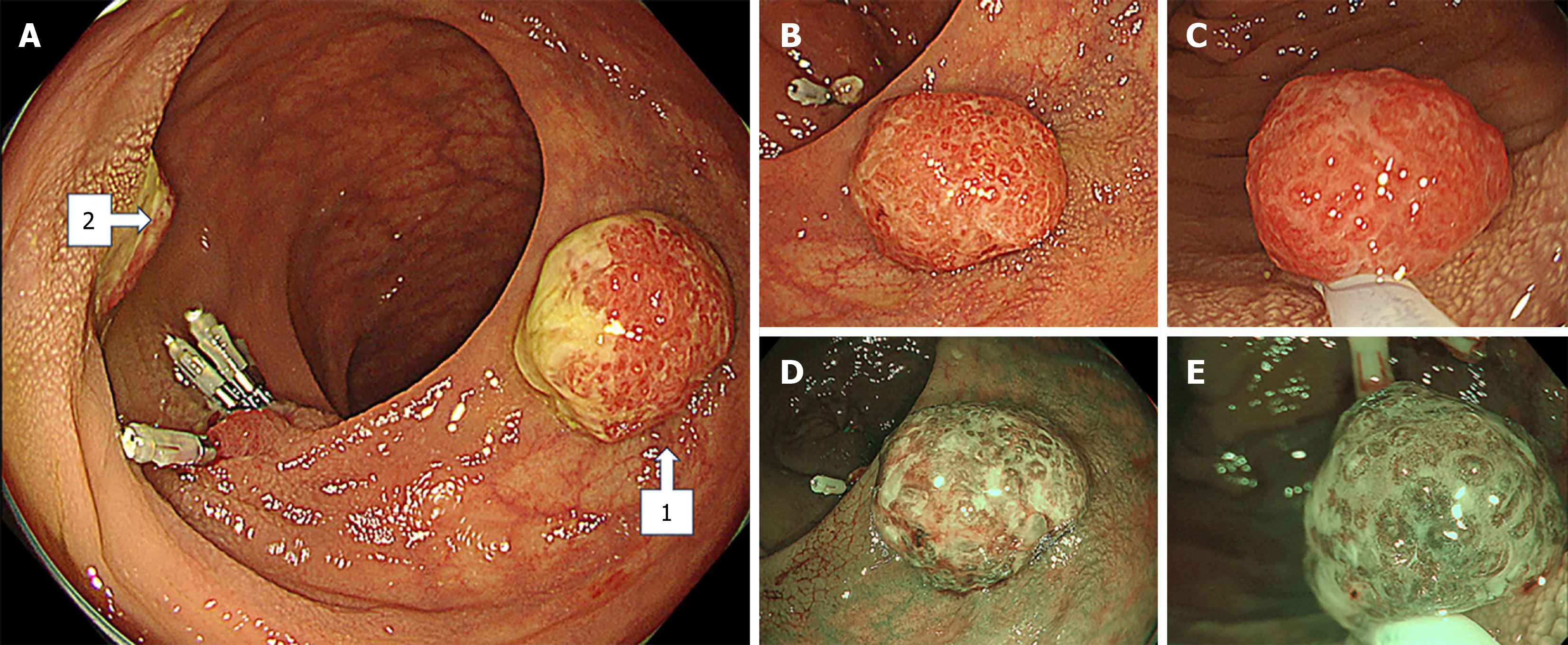
Figure 4 Semi-pedunculated polyps near the clipped bleeding site in the rectum.
A: Two semi-pedunculated polyps, approximately 15 mm in diameter (arrow 1 and arrow 2), are visible near the clipped bleeding site in the rectum; B and C: The polyps are intensely erythematous and have smooth surfaces and erosions with moss-white colorings; D and E: Narrow band imaging reveals dilated glands.
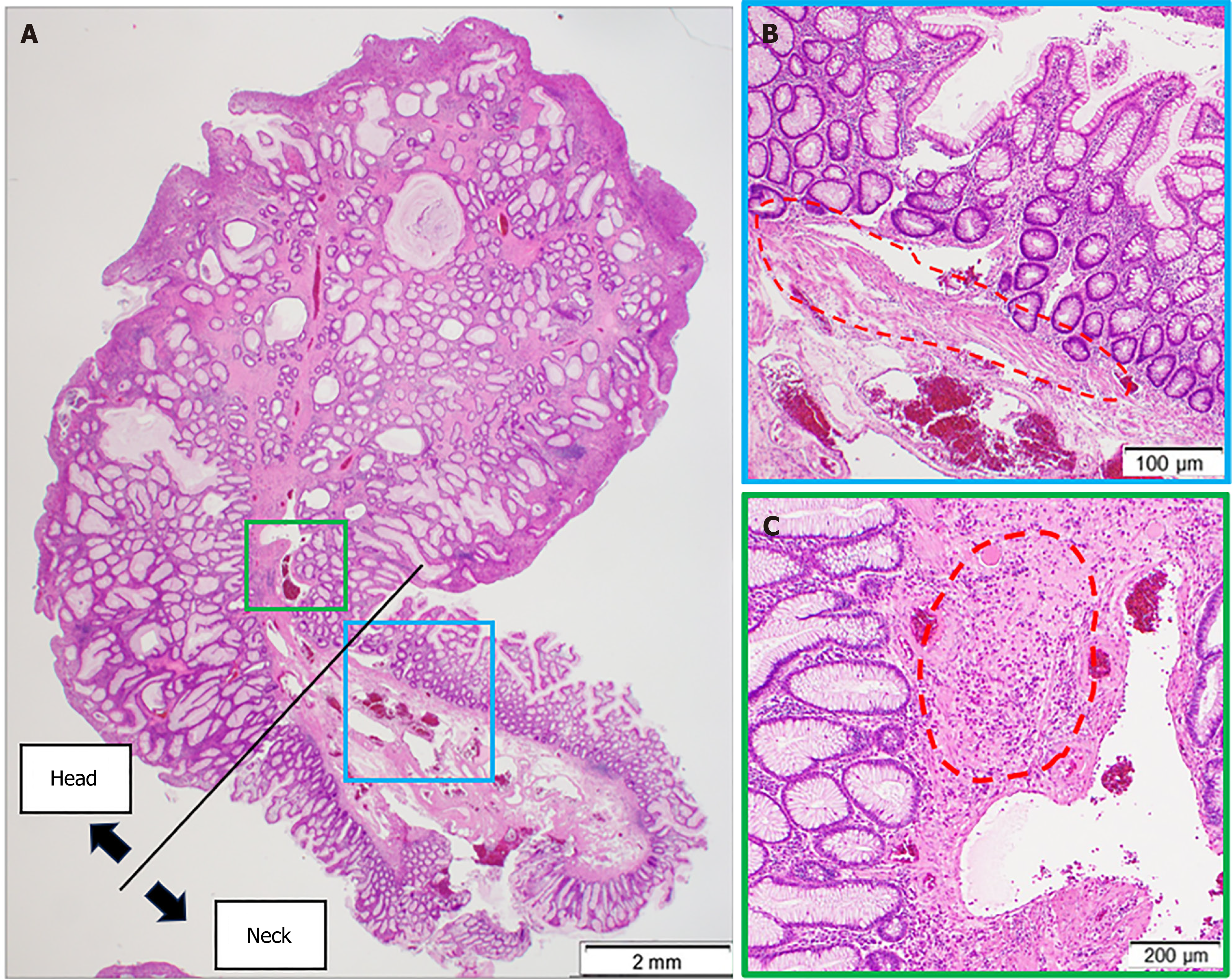
Figure 5 Histopathological evaluation of juvenile polyps.
A: Glandular ducts with dilated lumens distributed in the polyp; B: At the stem of the polyp (blue box in panel A, the lamina muscularis mucosae are present (dotted circle); C: Whereas in the head (green box in panel A), the lamina muscularis mucosae are lacking and a high inflammatory cell infiltrate is present (dotted circle).
- Citation: Kataoka F, Nakanishi T, Araki H, Ichino S, Kamei M, Makino H, Nagao R, Asano T, Tagami A, Moriwaki H. Adult juvenile polyp bleeding detected by extravascular contrast leakage and treated with endoscopic clipping: A case report. World J Gastrointest Endosc 2025; 17(2): 101135
- URL: https://www.wjgnet.com/1948-5190/full/v17/i2/101135.htm
- DOI: https://dx.doi.org/10.4253/wjge.v17.i2.101135


