Copyright
©The Author(s) 2025.
World J Gastrointest Endosc. Jan 16, 2025; 17(1): 102185
Published online Jan 16, 2025. doi: 10.4253/wjge.v17.i1.102185
Published online Jan 16, 2025. doi: 10.4253/wjge.v17.i1.102185
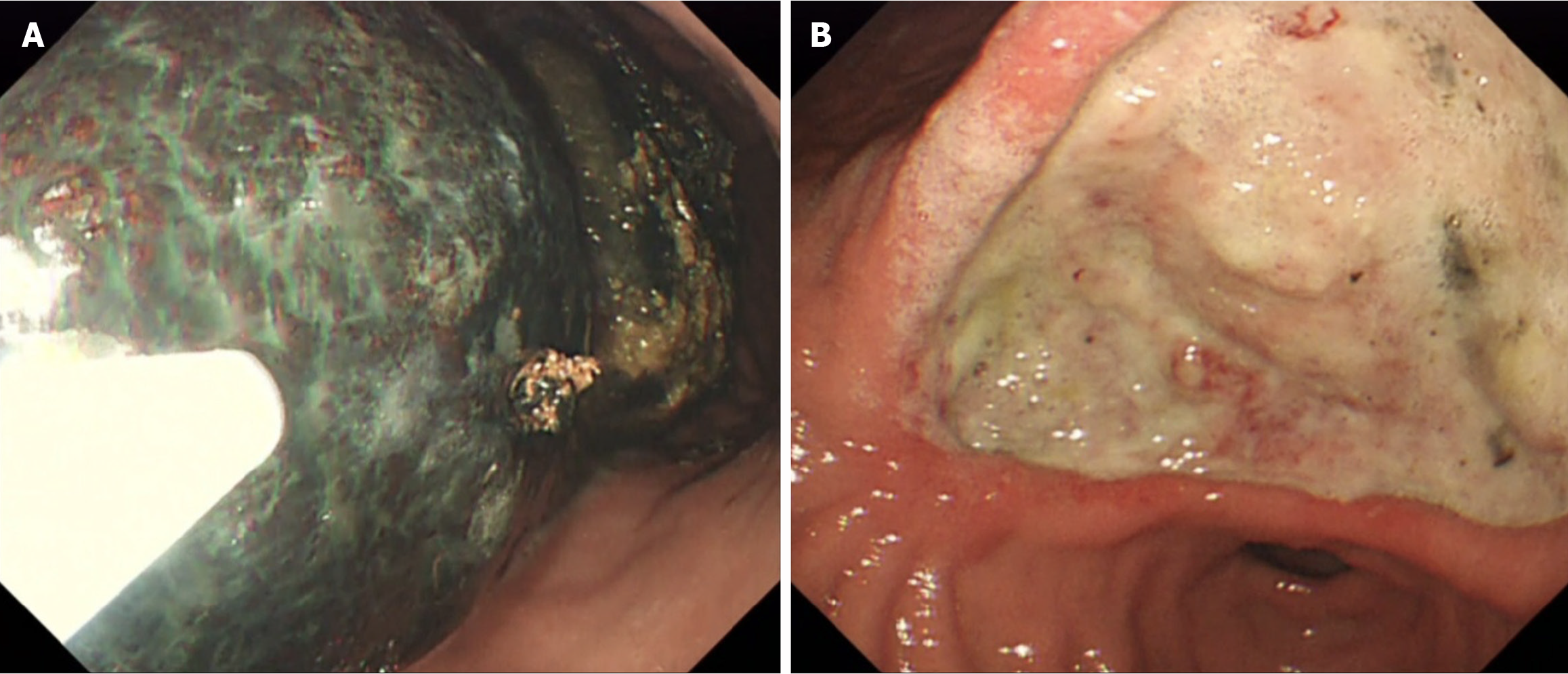
Figure 1 Esophagogastroduodenoscopy.
A: Two large phytobezoars, each 8 cm in diameter, were located in the fundus and body of the stomach; B: A Forrest IIb ulcer (5-6 cm in size) was located at the angular incisure and extended into the antrum area.
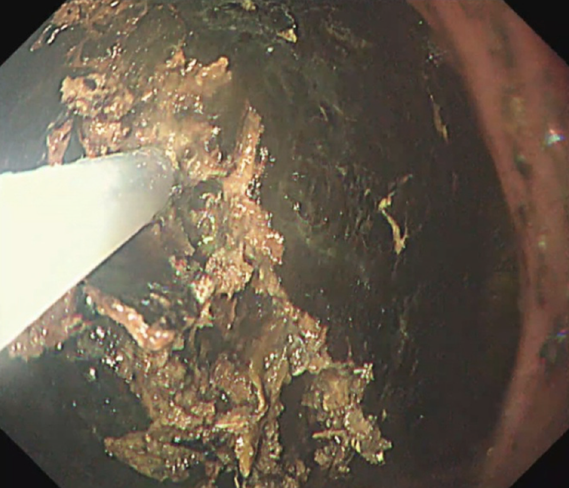
Figure 2
Snare-tip electrocautery successfully fragmented the bezoar.
- Citation: Lim CH, Lim CJ, Yao CT, Chang CC. Novel approach to managing two enormous bezoars with successive snare-tip electrocautery: A case report. World J Gastrointest Endosc 2025; 17(1): 102185
- URL: https://www.wjgnet.com/1948-5190/full/v17/i1/102185.htm
- DOI: https://dx.doi.org/10.4253/wjge.v17.i1.102185


