Copyright
©The Author(s) 2025.
World J Gastrointest Endosc. Jan 16, 2025; 17(1): 101534
Published online Jan 16, 2025. doi: 10.4253/wjge.v17.i1.101534
Published online Jan 16, 2025. doi: 10.4253/wjge.v17.i1.101534
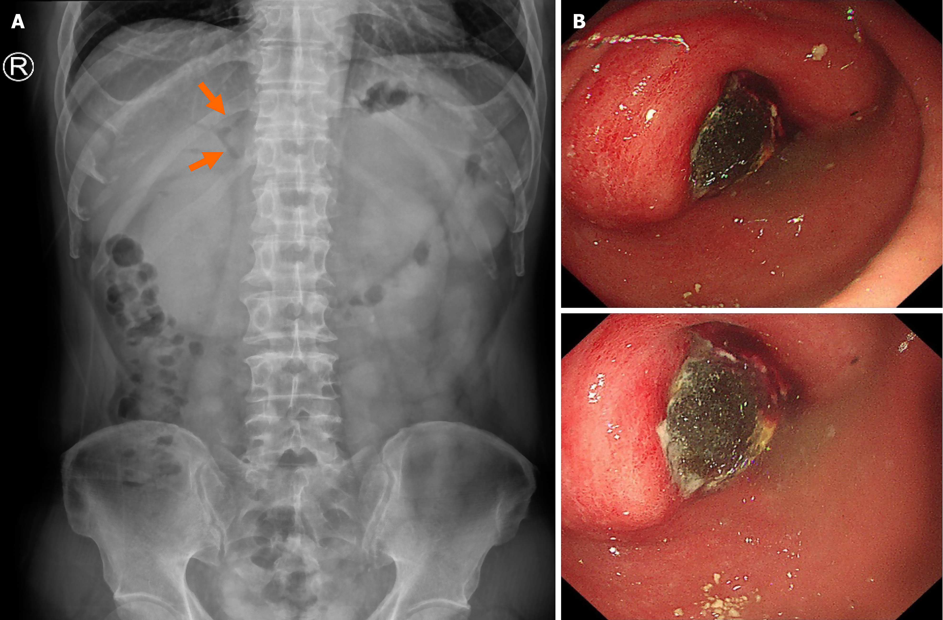
Figure 1 X-ray and Endoscopic findings.
A: Abdominal X-ray image showing pneumobilia of the intrahepatic and extrahepatic bile ducts (arrows); B: Endoscopy revealing a large gallstone in the gastric pylorus leading to gastric outlet obstruction.
Figure 2 Computed tomography.
A: Coronal computed tomography (CT) scan showing pneumobilia (asterisk) and a cholecystogastric fistula (arrowhead) with a large gallstone in the gastric antrum (arrows); B: Axial CT scan showing chronic cholecystitis with a gas-filled gallbladder and gallstones in the gastric antrum (arrow).
- Citation: Yang Y, Zhong DF. Cholecystogastric fistula presenting as pyloric obstruction - a Bouveret’s syndrome: A case report. World J Gastrointest Endosc 2025; 17(1): 101534
- URL: https://www.wjgnet.com/1948-5190/full/v17/i1/101534.htm
- DOI: https://dx.doi.org/10.4253/wjge.v17.i1.101534


