Copyright
©The Author(s) 2024.
World J Gastrointest Endosc. Aug 16, 2024; 16(8): 494-499
Published online Aug 16, 2024. doi: 10.4253/wjge.v16.i8.494
Published online Aug 16, 2024. doi: 10.4253/wjge.v16.i8.494
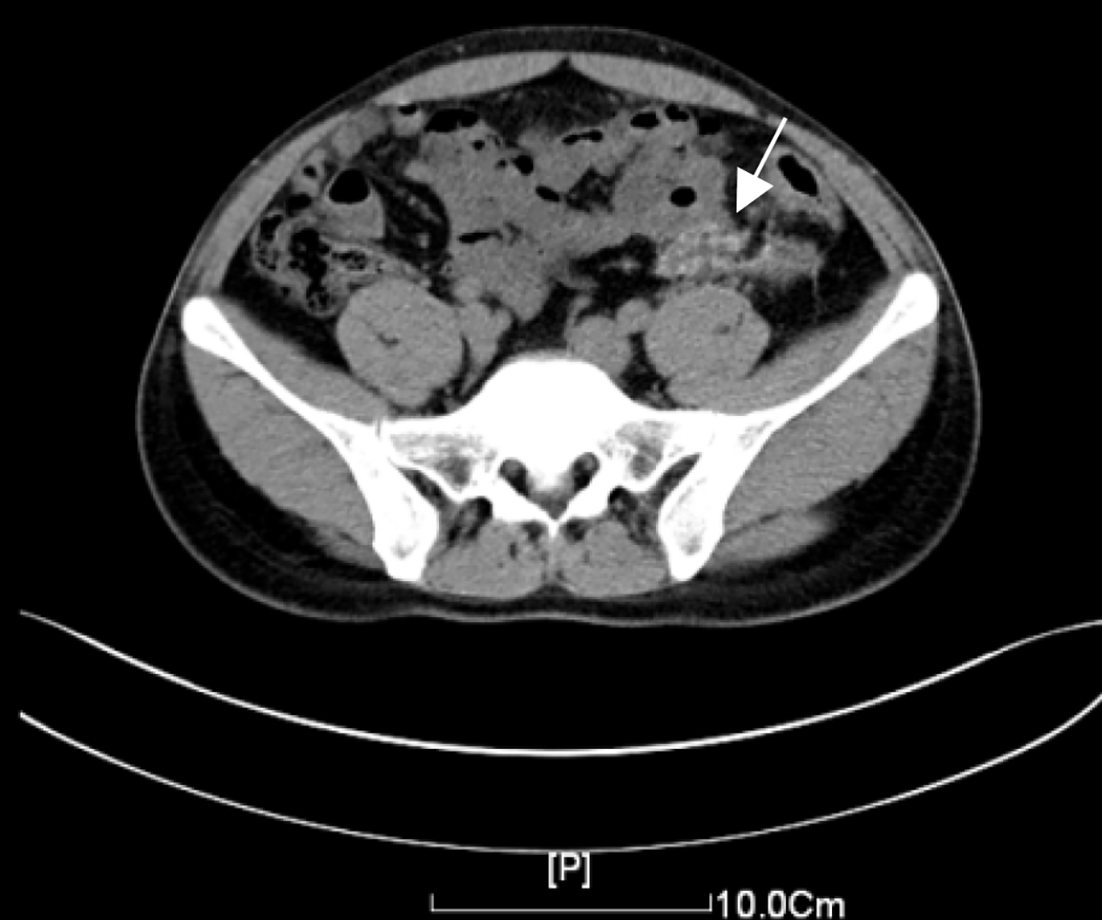
Figure 1 Computed tomography scan.
Image revealed irregular thickening of the left descending colon wall, luminal narrowing, an irregular patchy infiltrative density in the surrounding mesentery, and internal calcifications, measuring approximately 3.40 cm × 1.70 cm in cross-sectional size.
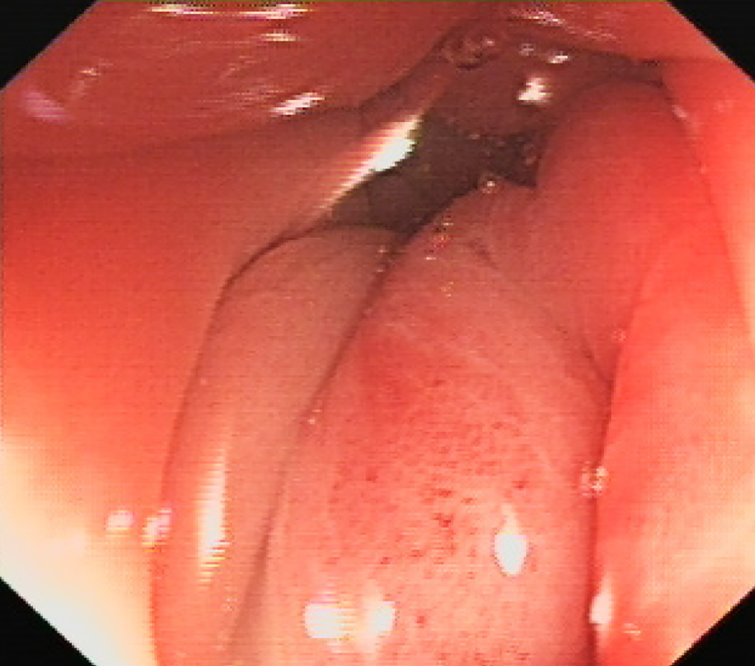
Figure 2 Colonoscopy findings.
Colonoscopy revealed luminal narrowing in the sigmoid colon segment, with highly congested and edematous mucosa.
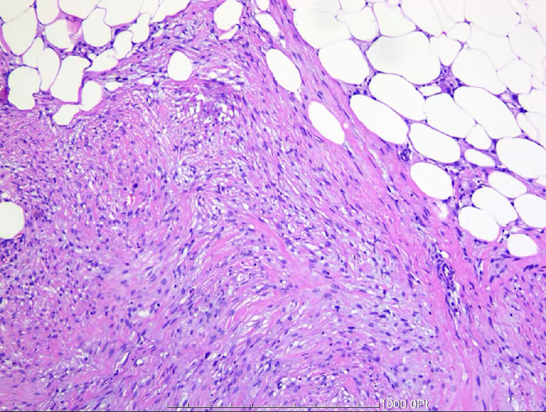
Figure 3 Postoperative pathological examination of tissue specimens.
Surgical pathology findings revealed spindle-shaped cells and adipocytes distributed in a patchy pattern within the tough area of the outer mesentery of the colonic wall, with spindle cells arranged in bundles or interlaced (some in a disordered manner). The cells had short spindle-shaped nuclei, which were slightly pointed at both ends, and stained cytoplasm. These findings suggested focal new bone formation, which was diagnosed as heterotopic mesenteric ossification.
- Citation: Zhang BF, Liu J, Zhang S, Chen L, Lu JZ, Zhang MQ. Heterotopic mesenteric ossification caused by trauma: A case report. World J Gastrointest Endosc 2024; 16(8): 494-499
- URL: https://www.wjgnet.com/1948-5190/full/v16/i8/494.htm
- DOI: https://dx.doi.org/10.4253/wjge.v16.i8.494


