Copyright
©The Author(s) 2024.
World J Gastrointest Endosc. Aug 16, 2024; 16(8): 472-482
Published online Aug 16, 2024. doi: 10.4253/wjge.v16.i8.472
Published online Aug 16, 2024. doi: 10.4253/wjge.v16.i8.472
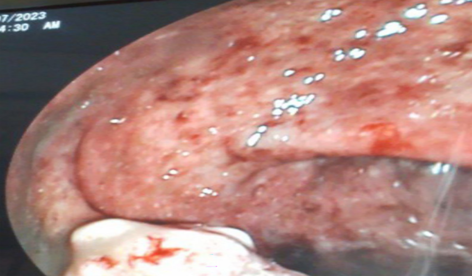
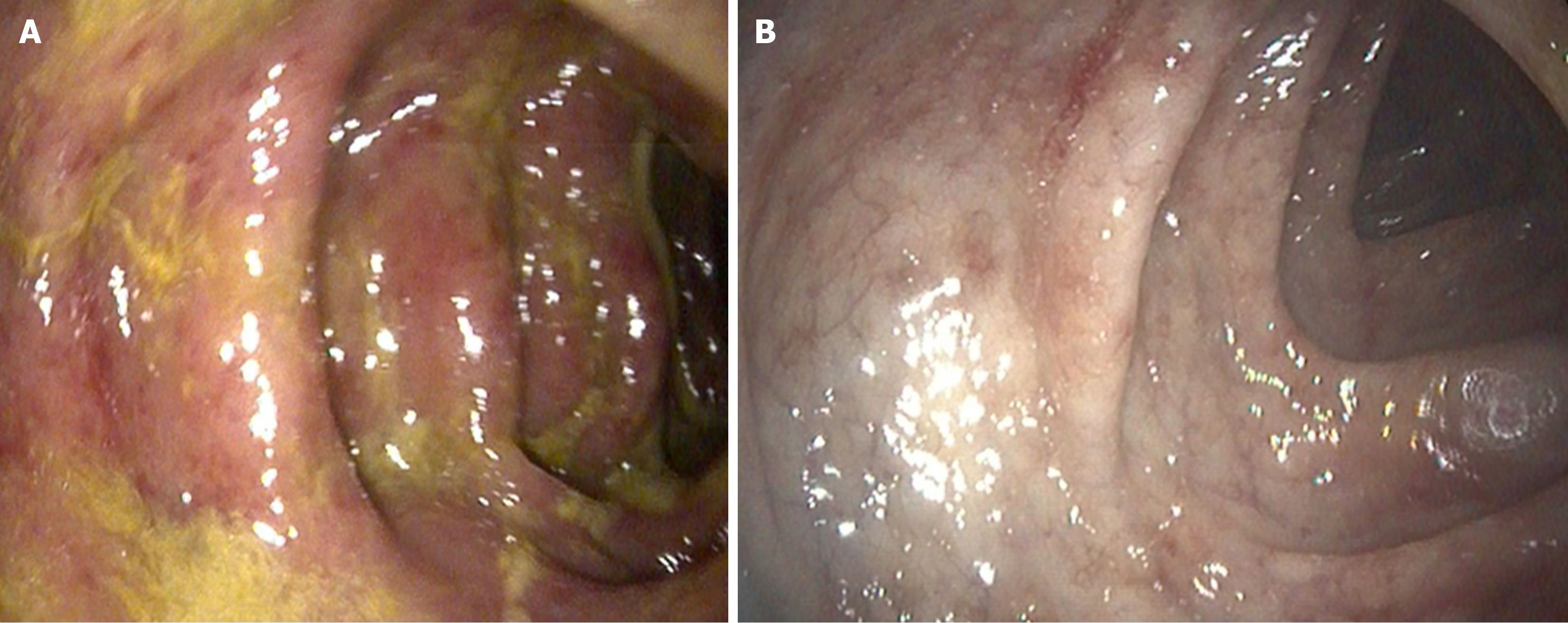
Figure 1 Colonoscopy image showing diffusely inflamed rectal mucosa and a mass-like lesion (Case 1).
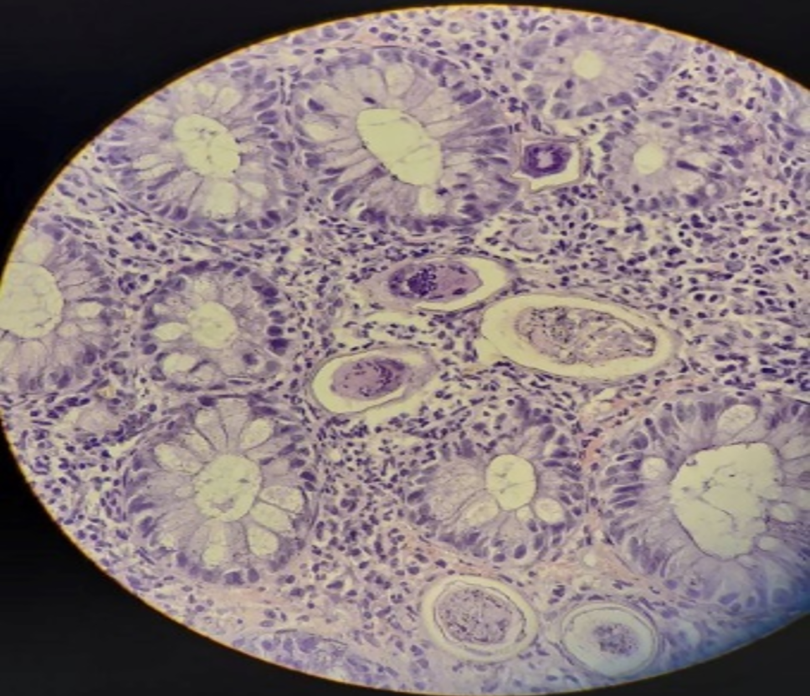
Figure 2 Biopsy showing numerous Schistosoma eggs (arrow) and chronic inflammatory infiltrates in the colonic mucosa for Case 1.
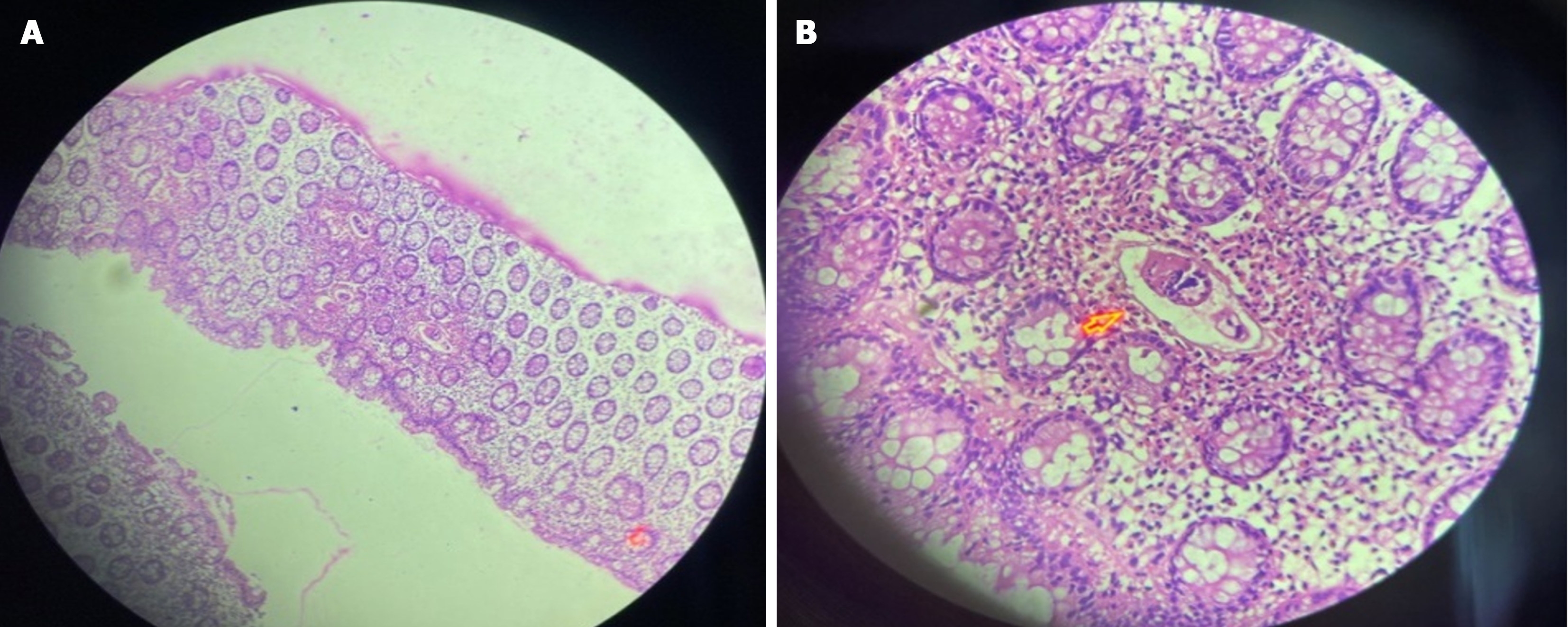
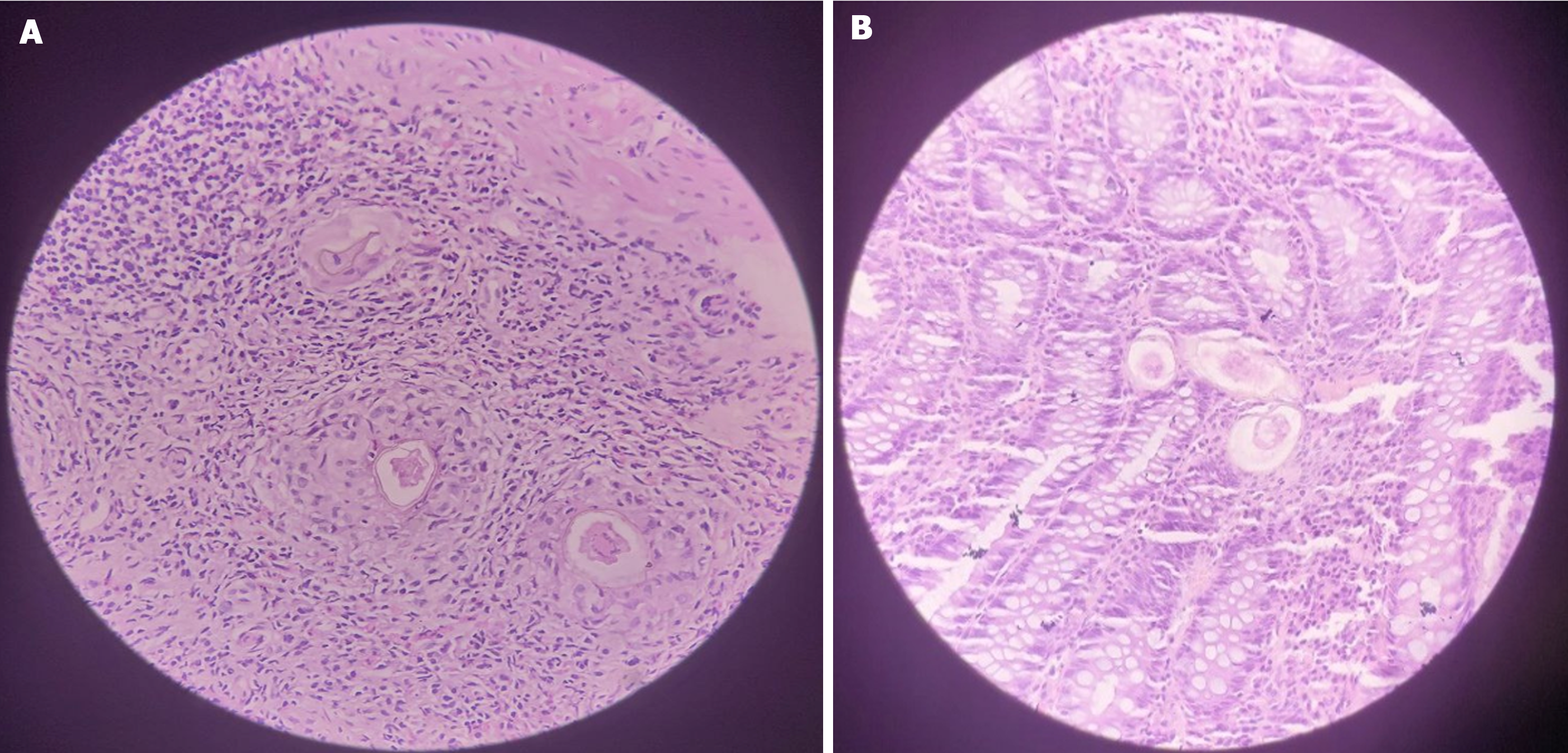
Figure 3 Microscope images.
A: Low power magnification photomicrographs showing Schistosoma eggs embedded within the mucosa surrounded by dense eosinophilic infiltrates (× 10 magnification); B: High power magnification of Schistosoma egg and surrounding eosinophil (× 40 magnification) for Case 2.
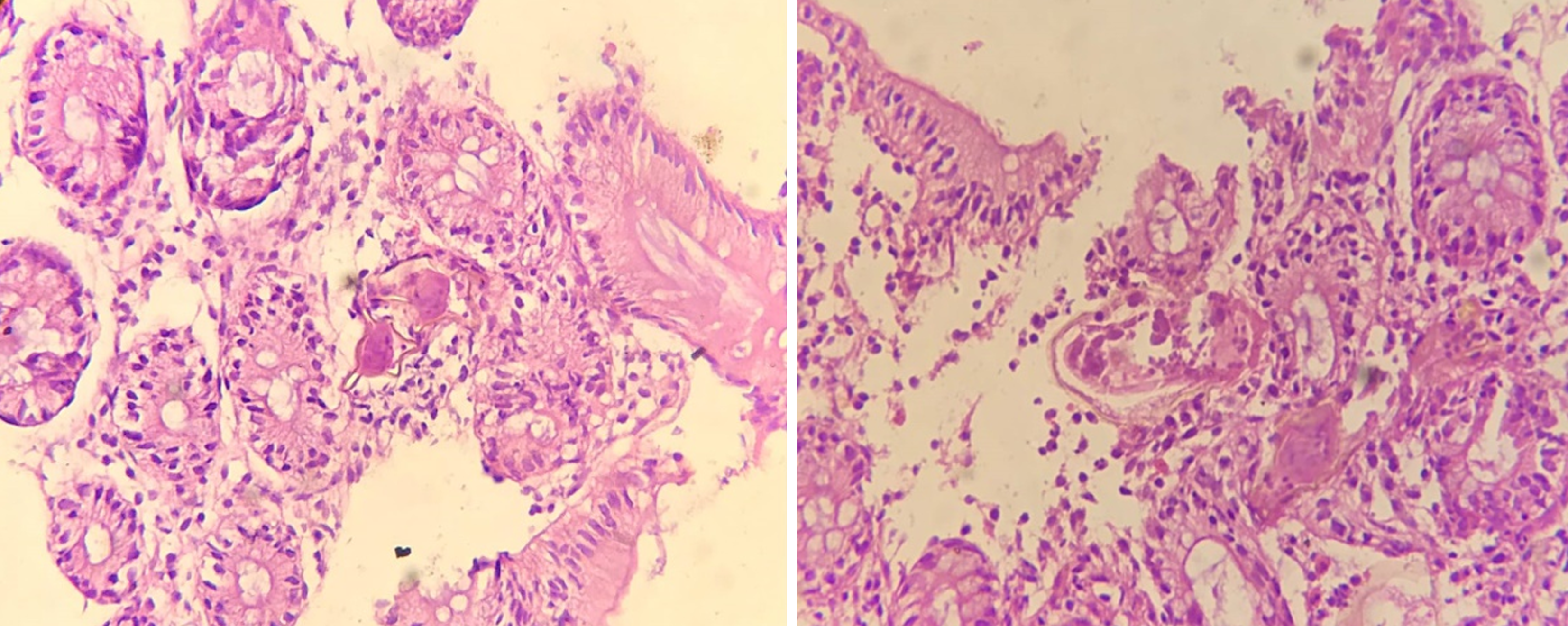
Figure 4 Schistosoma egg embedded within colonic mucosa, with a lateral spine with surrounding eosinophil predominant inflammatory response (× 40 magnification) for Case 3.
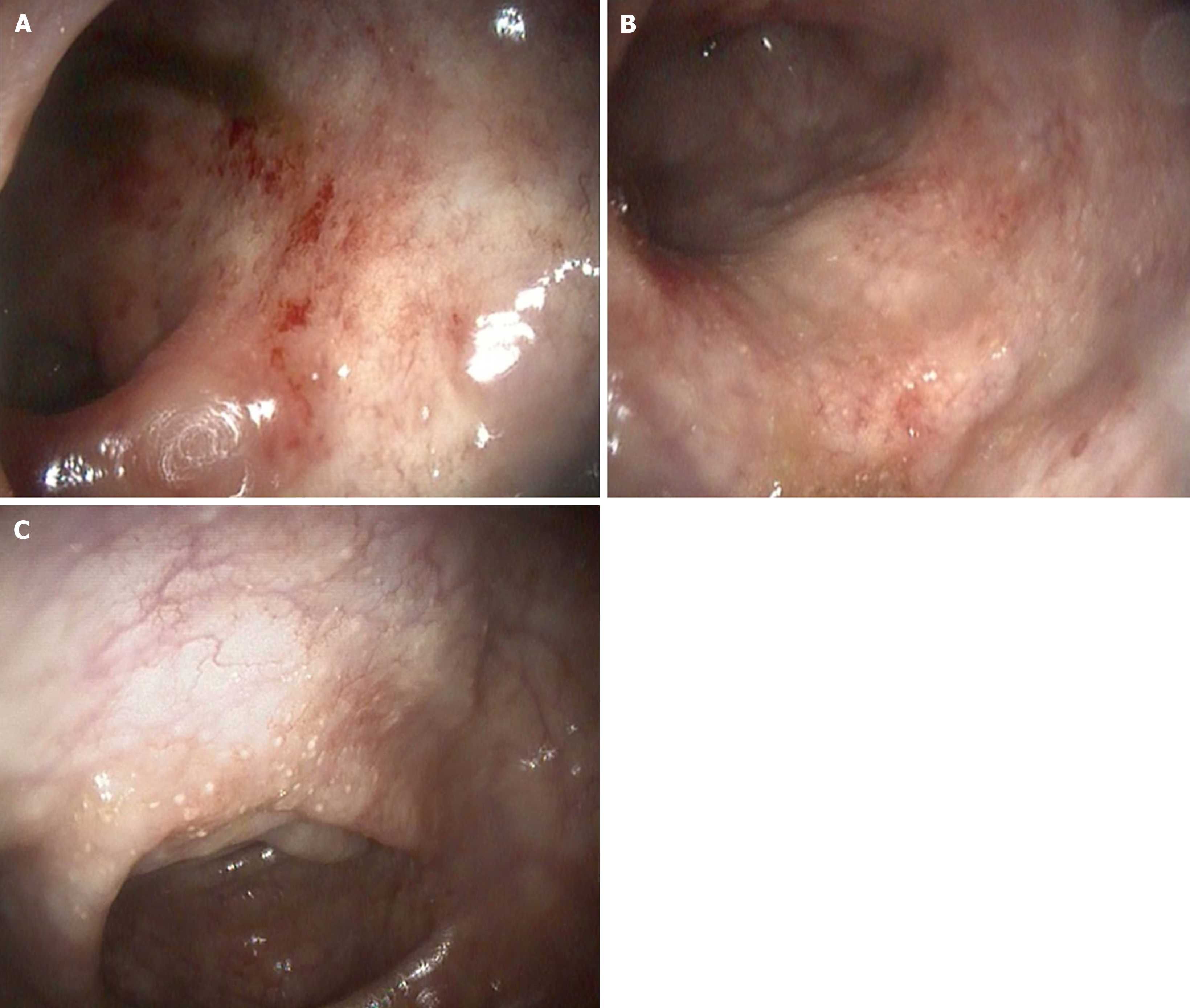
Figure 5 Patchy inflamed colonic mucosa (Case 4).
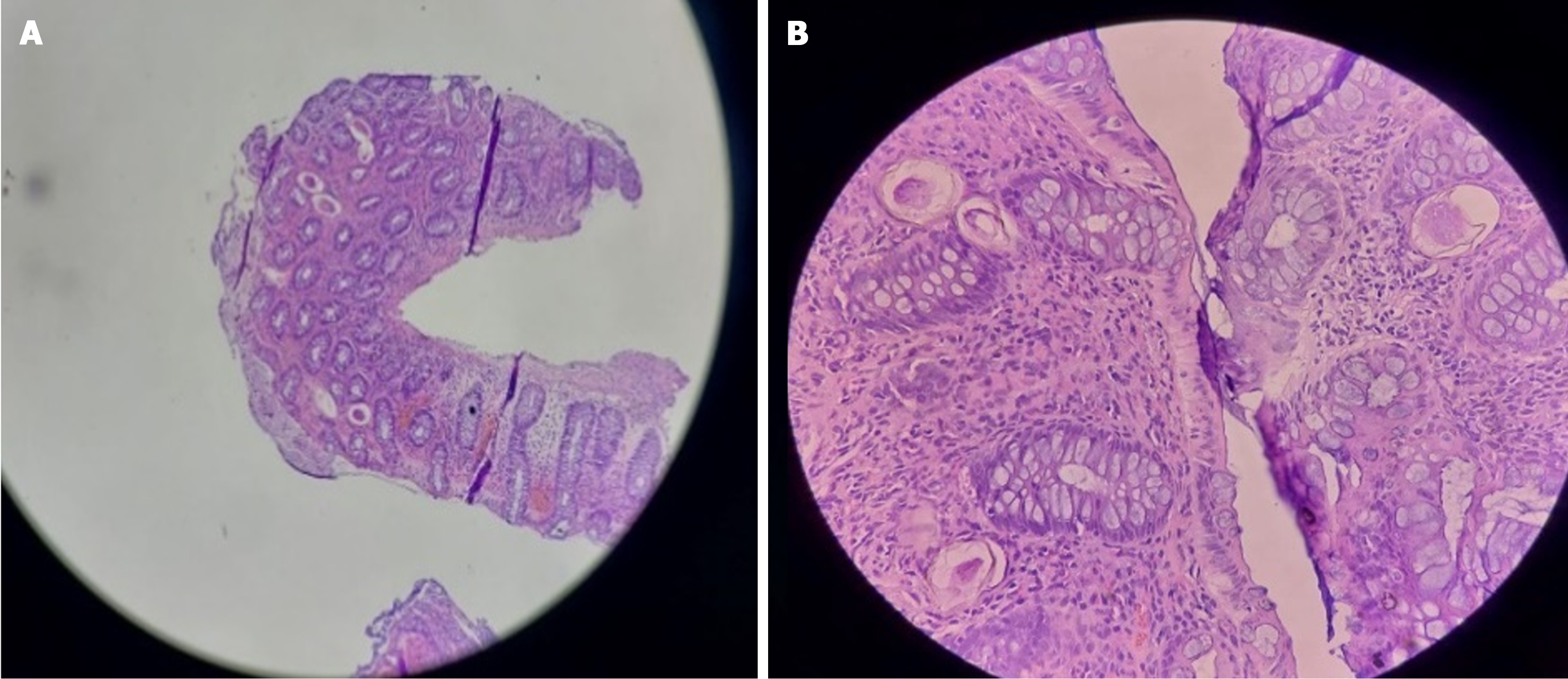
Figure 6 Several Schistosoma ova surrounded by mixed inflammatory cells predominated by eosinophils and with granuloma formation (Case 4).
Figure 7 Inflamed mucosa with granularity on colonoscopy (for Case 5).
Figure 8 Several Schistosoma eggs surrounded by inflammatory cells (Case 5).
- Citation: Mulate ST, Nur AM, Tasamma AT, Annose RT, Dawud EM, Ekubazgi KW, Mekonnen HD, Mohammed HY, Hailemeskel MB, Yimer SA. Colonic schistosomiasis mimicking cancer, polyp, and inflammatory bowel disease: Five case reports and review of literature. World J Gastrointest Endosc 2024; 16(8): 472-482
- URL: https://www.wjgnet.com/1948-5190/full/v16/i8/472.htm
- DOI: https://dx.doi.org/10.4253/wjge.v16.i8.472


