Copyright
©The Author(s) 2024.
World J Gastrointest Endosc. Jun 16, 2024; 16(6): 368-375
Published online Jun 16, 2024. doi: 10.4253/wjge.v16.i6.368
Published online Jun 16, 2024. doi: 10.4253/wjge.v16.i6.368
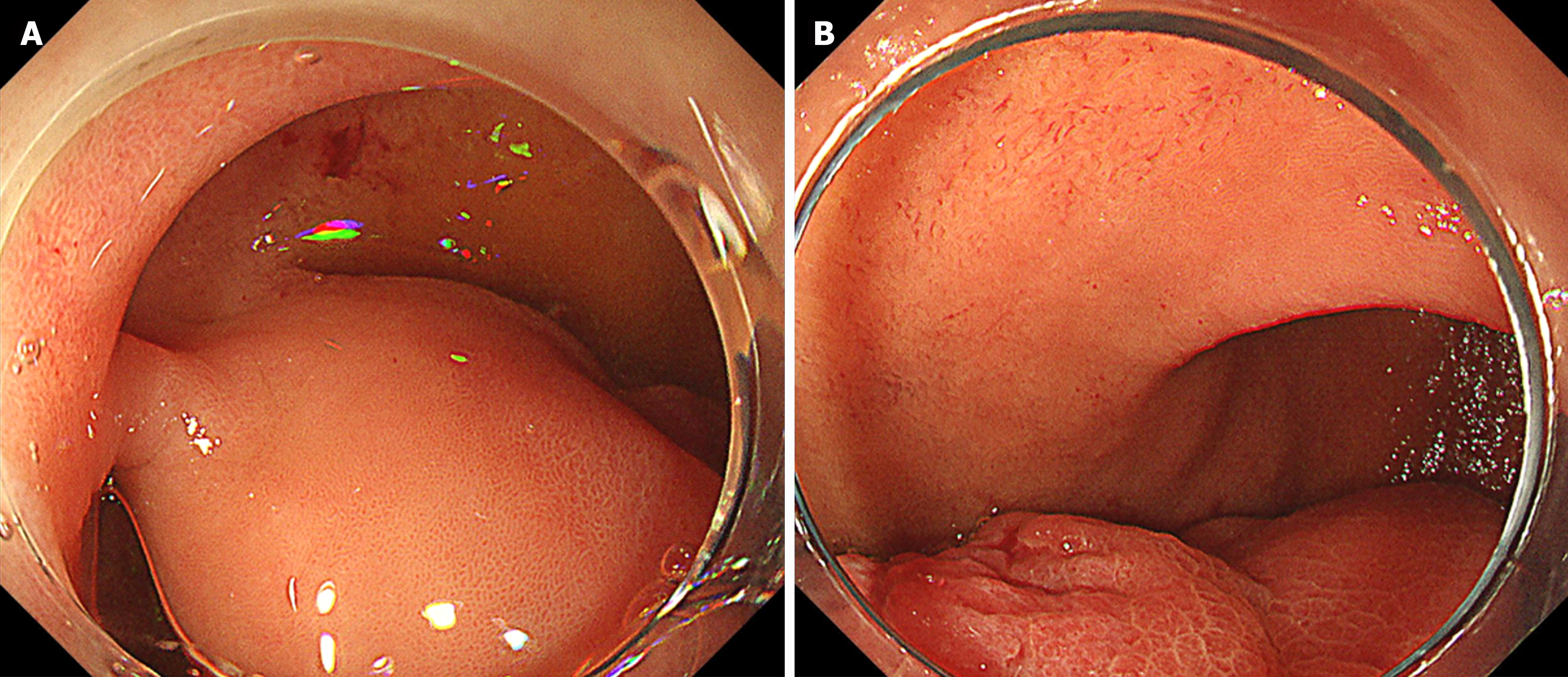
Figure 1 Endoscopic images of the duodenal lesion.
A: Endoscopic image of the base of the lesion; B: Endoscopic image of the middle to the tip of the lesion.
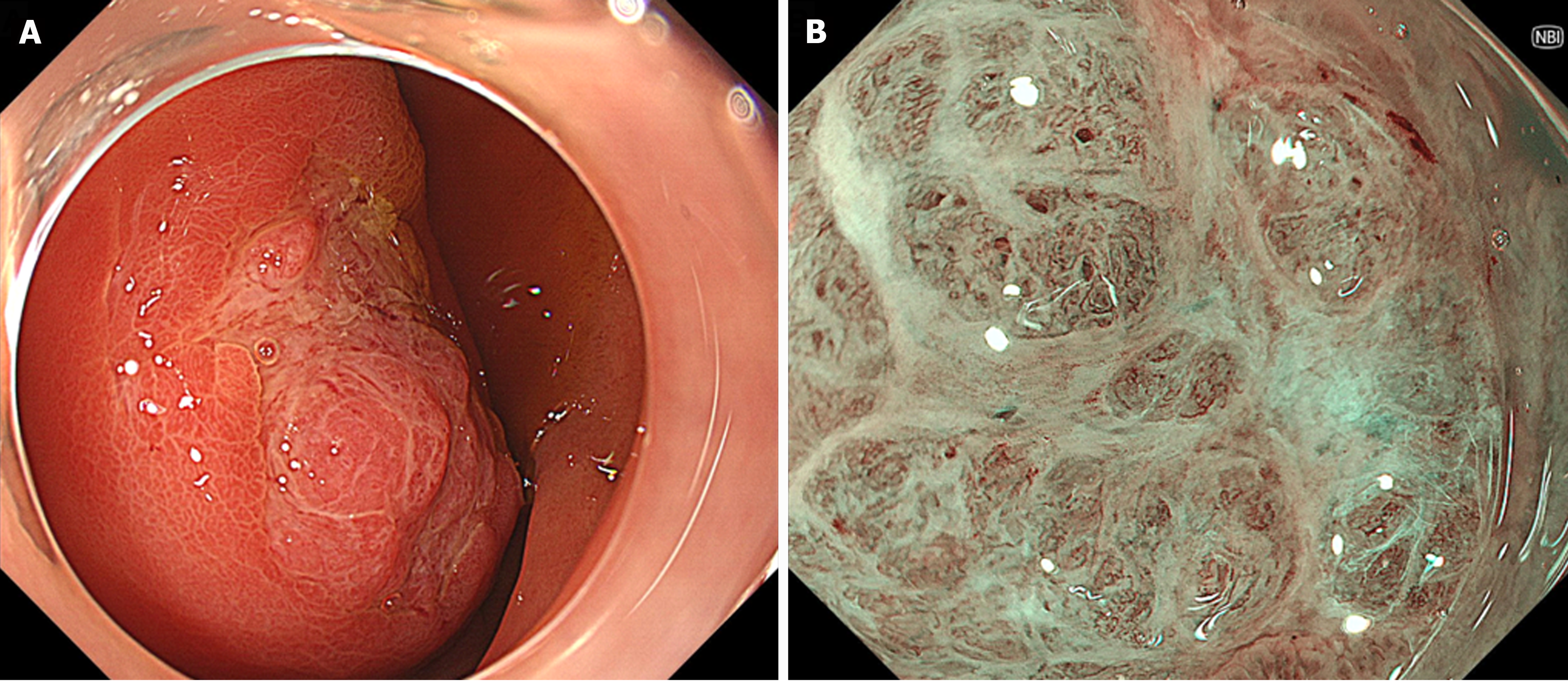
Figure 2 A depression with a mucosal defect in the lesion.
A: In a portion of the lesion, a depression with a mucosal defect was seen; B: Magnified narrow-band imaging observation of this depressed area revealed structures resembling small round glandular openings and band-like septum-like structures suggestive of fibrous components.
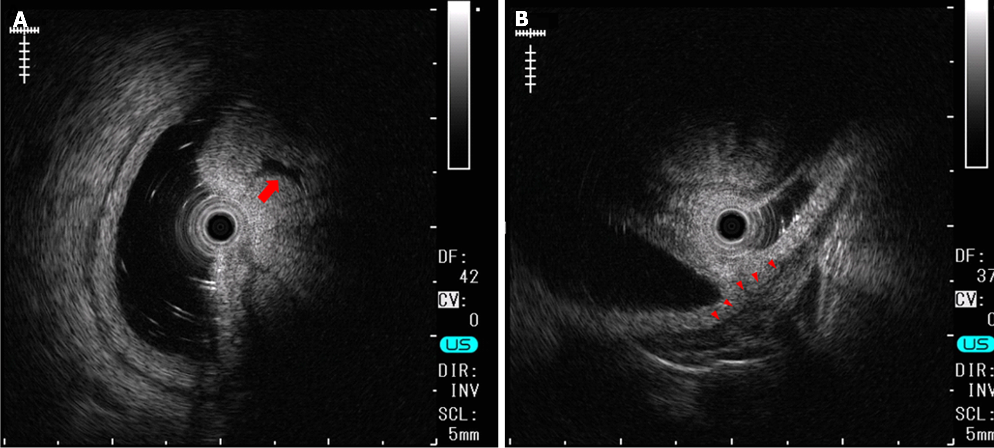
Figure 3 Endoscopic ultrasonography.
A: Endoscopic ultrasonography revealed that the lesion had relatively high echogenicity with a cystic component (arrow) inside; B: At the base of the lesion, a slight elevation of a low echogenicity layer that was suggestive of the muscular layer was seen (arrowhead).
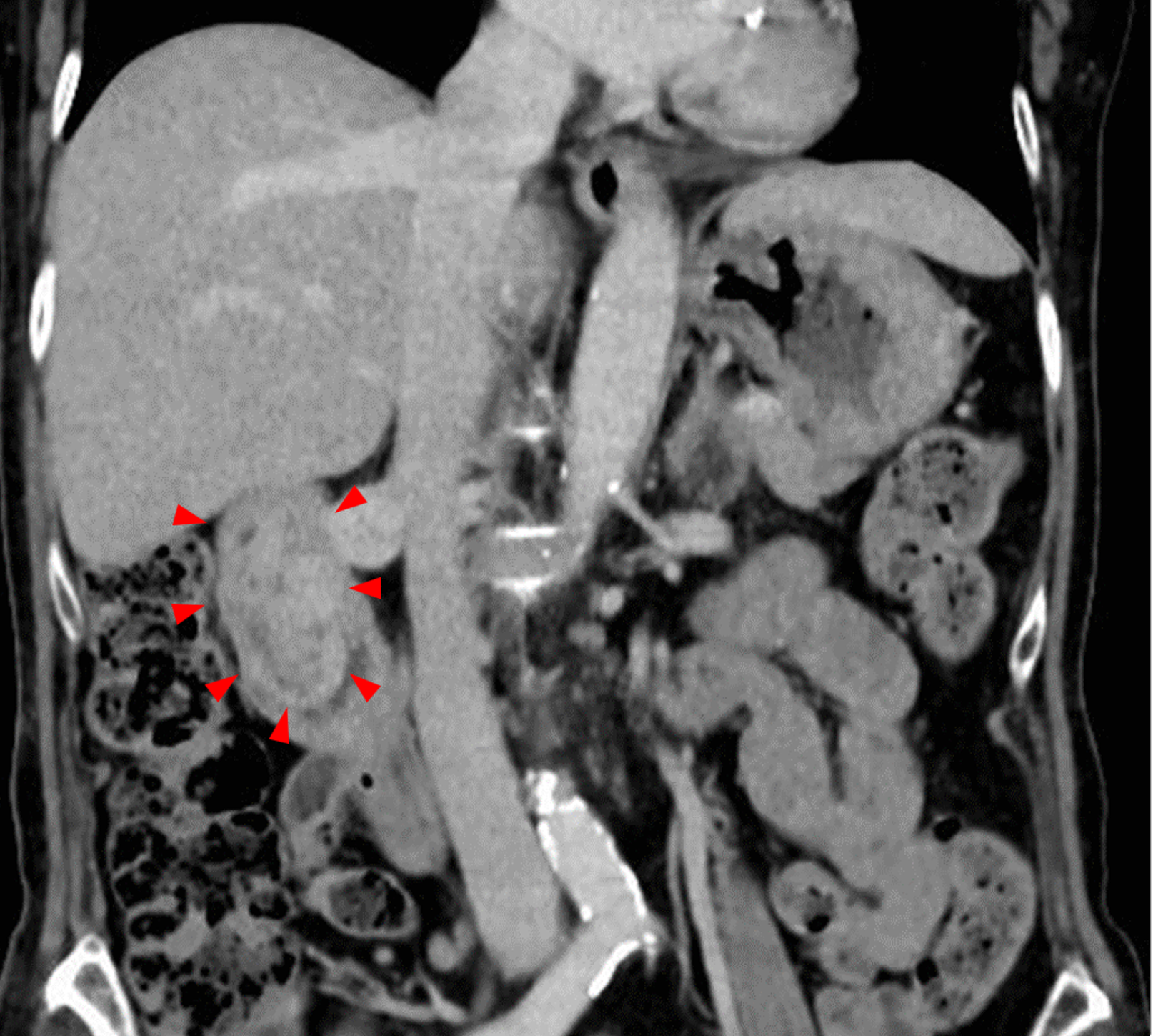
Figure 4 Abdominal contrast-enhanced computed tomography.
It revealed a 7 cm protruding lesion (arrowhead) hanging from the bulb to the descending portion of the duodenum. The lesion showed minimal enhancement in the arterial phase and a mosaic-like enhancement pattern in the venous phase.
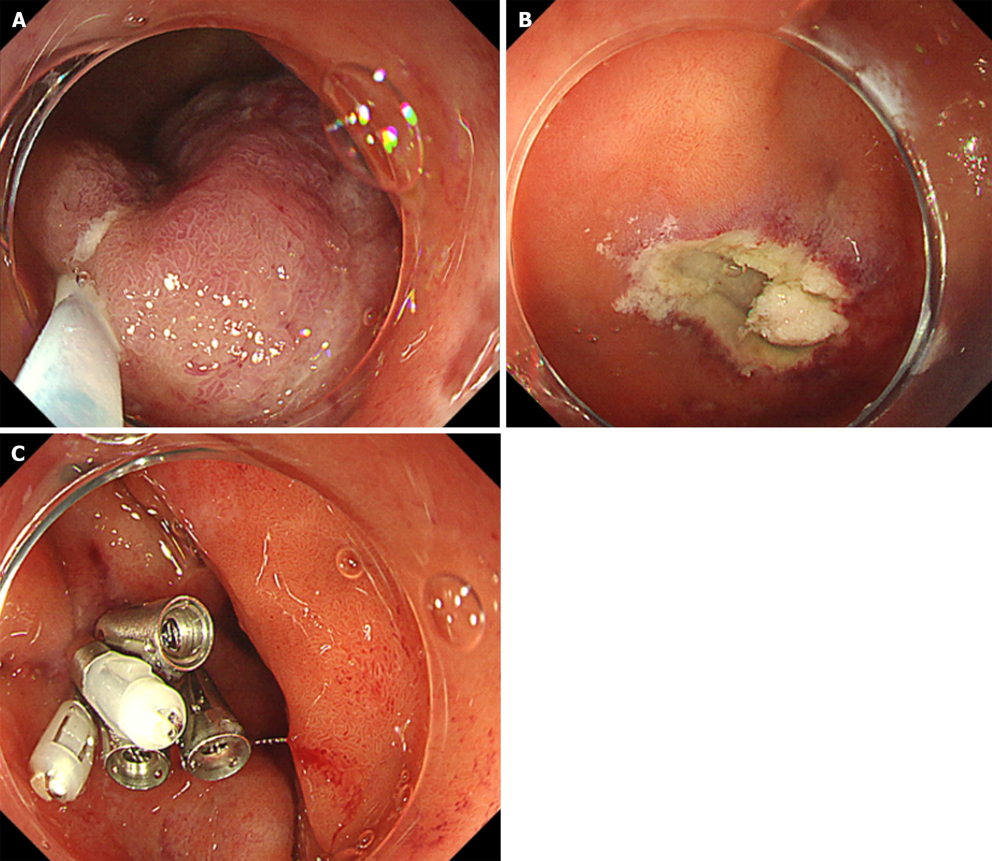
Figure 5 Endoscopic mucosal resection of the lesion.
A: After injection of hypertonic saline epinephrine solution at the base of the lesion, we grasped it with a snare. We attempted to excise the lesion using the cutting mode of the high-frequency generator. There was no electrical activation the first time. After relocating the constriction site with the snare slightly away from the base, we achieved satisfactory electrical conduction, and the lesion was excised entirely; B: There was no macroscopic residual tumor on the ulcerated surface after excising the lesion, and there were no muscle layer injuries or perforations; C: The ulceration after excision was completely closed using endoclips.
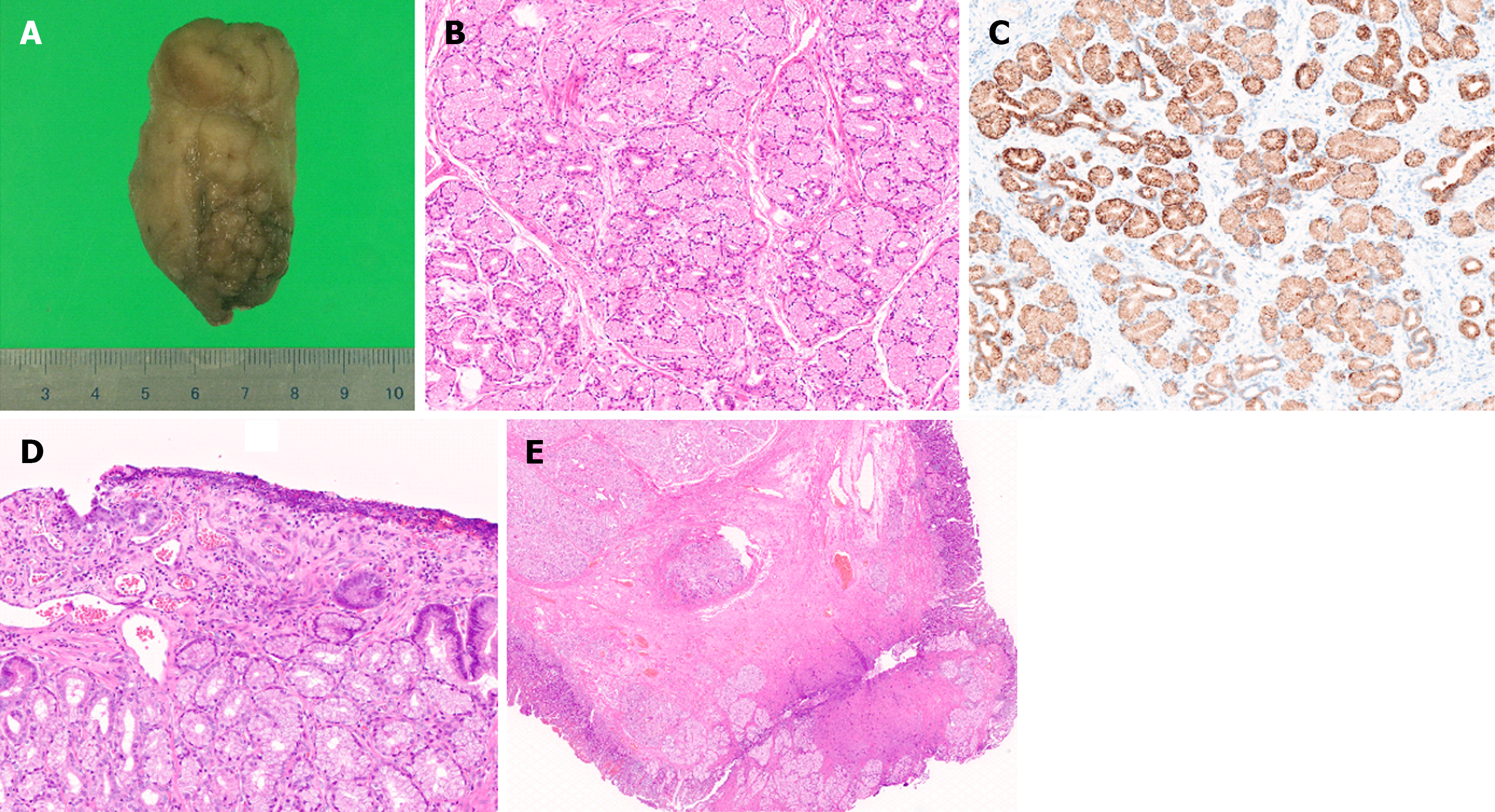
Figure 6 Pathological findings of the lesion.
A: The lesion size after formalin fixation was 6 cm × 3.5 cm × 3 cm; B: Histopathological examination revealed nodular proliferation of Brunner's glands without atypia, partitioned by fibrous septa; C: This site exhibited positive staining for MUC6; D: The area observed as a depressed region during EGD showed superficial erosion and regenerating epithelium; E: At the resection margin, thickened muscularis mucosa, fibrosis in the submucosal layer, and normal Brunner's glands were observed.
- Citation: Makazu M, Sasaki A, Ichita C, Sumida C, Nishino T, Nagayama M, Teshima S. Giant Brunner's gland hyperplasia of the duodenum successfully resected en bloc by endoscopic mucosal resection: A case report. World J Gastrointest Endosc 2024; 16(6): 368-375
- URL: https://www.wjgnet.com/1948-5190/full/v16/i6/368.htm
- DOI: https://dx.doi.org/10.4253/wjge.v16.i6.368


