Copyright
©The Author(s) 2024.
World J Gastrointest Endosc. Jun 16, 2024; 16(6): 305-317
Published online Jun 16, 2024. doi: 10.4253/wjge.v16.i6.305
Published online Jun 16, 2024. doi: 10.4253/wjge.v16.i6.305
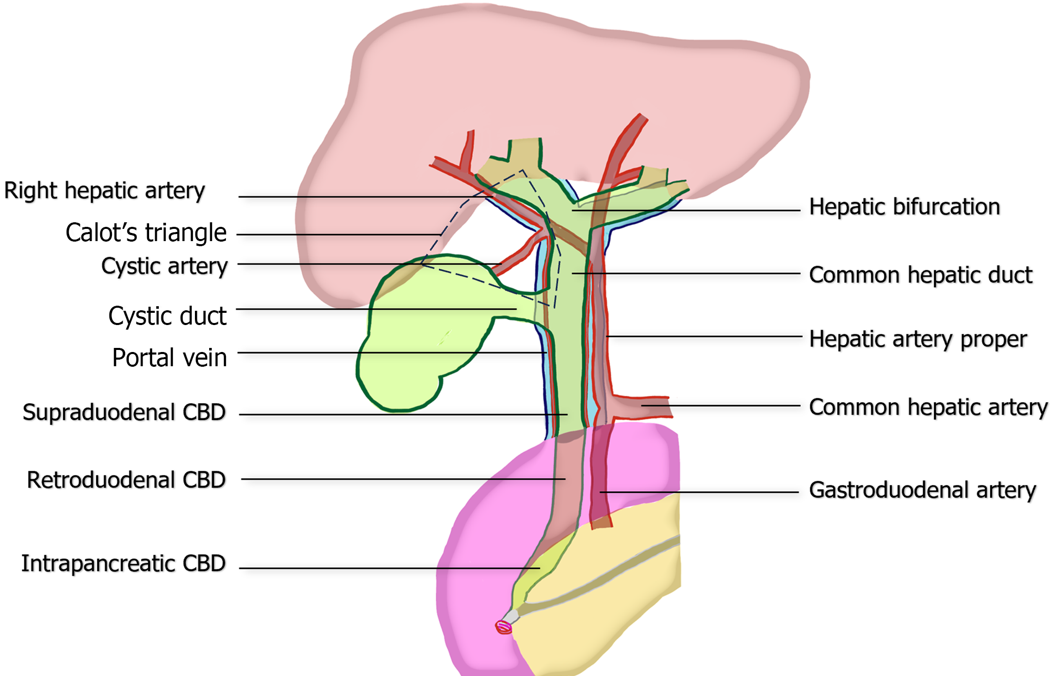
Figure 1 Common anatomy of portal triad, illustrating the relationship between the extrahepatic bile duct, hepatic artery proper, and portal vein.
The figure also demonstrates the boundaries of the hepatocystic triangle of Calot and vascular supply to common bile duct at the 3 and 9 o’clock position. CBD: Common bile duct
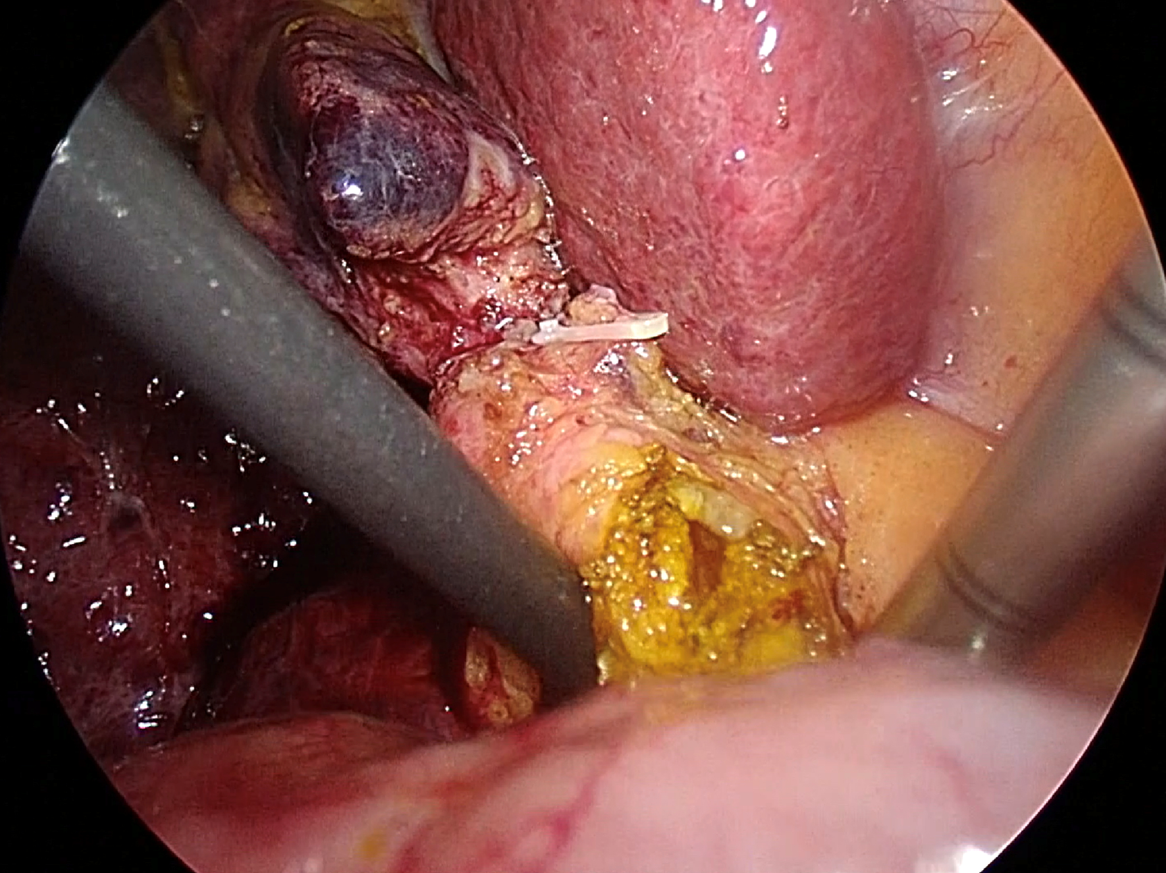
Figure 2 Transductal bile duct stone removal using the chopstick technique.
Two atraumatic instruments are placed on the right-posterior and the left-anterior side of the common bile duct.
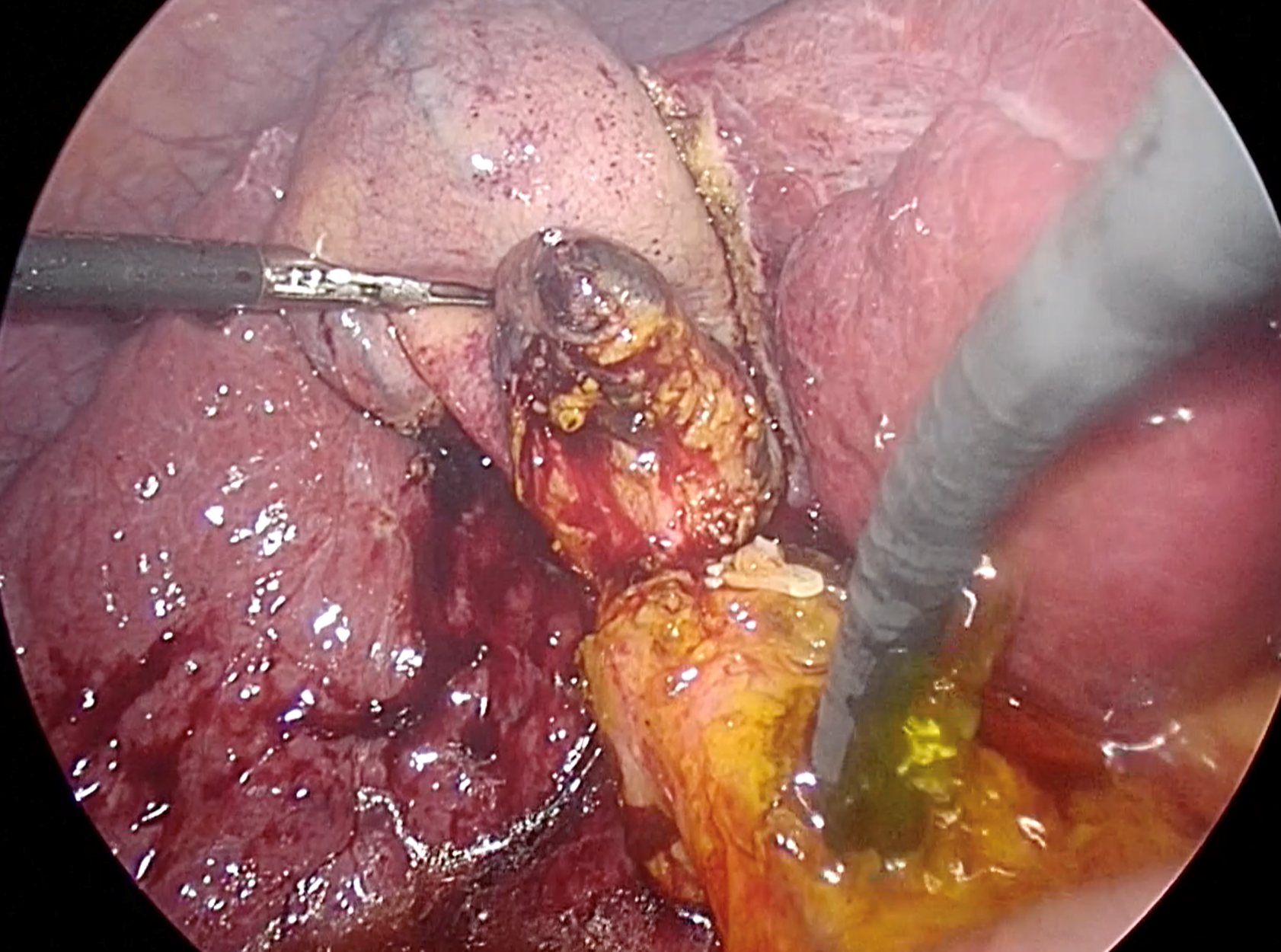
Figure 3 Confirmation of complete ductal clearance by choledochoscopy.
The choledochoscope should pass proximally to visualize the left and right intrahepatic ducts, as well as distally to the duodenum, to ensure complete ductal examination. Additionally, irrigation with normal saline solution via the choledochoscope proves beneficial for flushing out small stones and sludge.
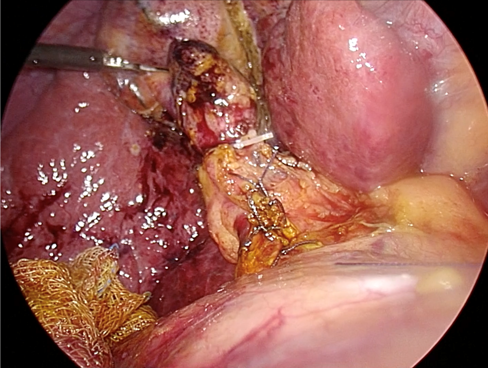
Figure 4 Ductal closure following complete transductal common bile duct stone removal.
Typically, intracorporeal suturing with interrupted 4-0 or 5-0 absorbable sutures is utilized. Based on current evidence, the closure-over-stent technique, which involves inserting a 7-10 French biliary stent followed by primary suture, is recommended.
- Citation: Suwatthanarak T, Chinswangwatanakul V, Methasate A, Phalanusitthepha C, Tanabe M, Akita K, Akaraviputh T. Surgical strategies for challenging common bile duct stones in the endoscopic era: A comprehensive review of current evidence. World J Gastrointest Endosc 2024; 16(6): 305-317
- URL: https://www.wjgnet.com/1948-5190/full/v16/i6/305.htm
- DOI: https://dx.doi.org/10.4253/wjge.v16.i6.305


