Copyright
©The Author(s) 2024.
World J Gastrointest Endosc. Oct 16, 2024; 16(10): 557-565
Published online Oct 16, 2024. doi: 10.4253/wjge.v16.i10.557
Published online Oct 16, 2024. doi: 10.4253/wjge.v16.i10.557
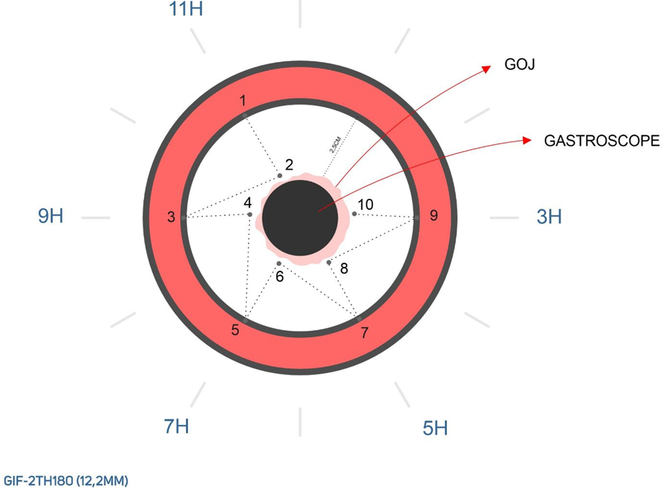
Figure 1
Diagram of suture placement in the fundus at the 1, 3, 5, 7, and 11 o’clock positions.
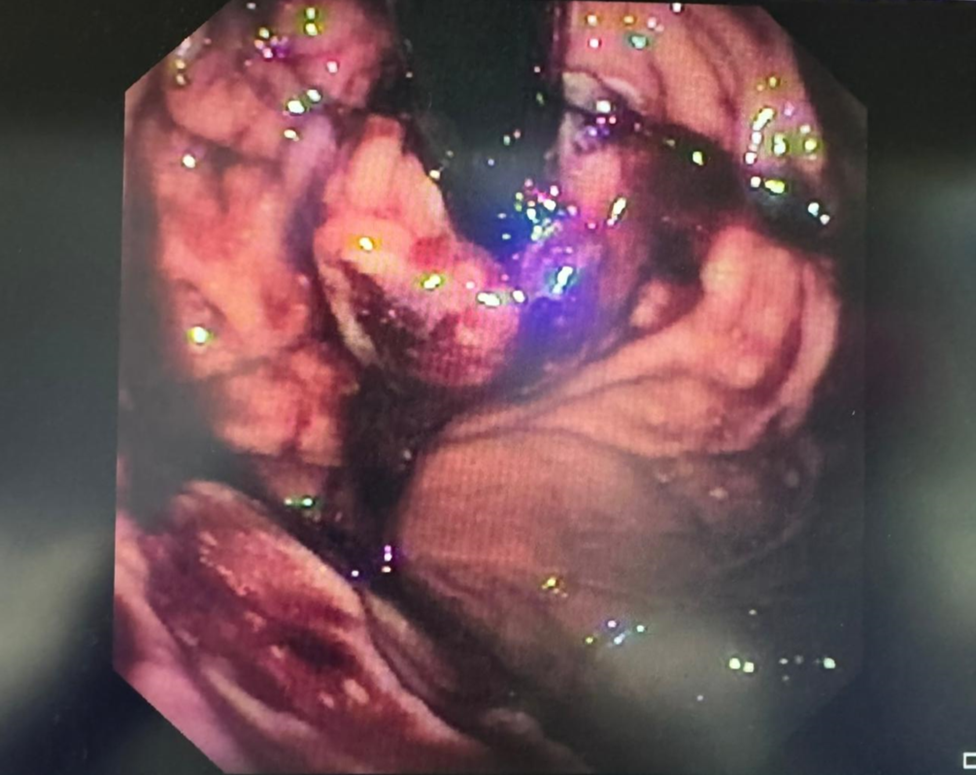
Figure 2
Endoscopic view after the placement of sutures in the fundus at 1, 3, 5, 7, and 11 o’clock positions.
Figure 3 Comparisons of gastroesophageal reflux disease-related quality of life and DeMeester scores at the time of the procedure, 6 months and 12 months after the procedure.
A: Mean gastroesophageal reflux disease (GERD)-related quality of life (QOL) (50) score at the time of the procedure, 6 and 12 months postoperatively; B: Mean GERD-related QOL (30) score was highest at the time of the procedure, followed by those at 6 and 12 months postoperatively (P < 0.001); C: Mean DeMeester score was highest at the time of the procedure, followed by those at 6 and 12 months postoperatively (P < 0.001). GERD: Gastroesophageal reflux disease; QOL: Quality of life.
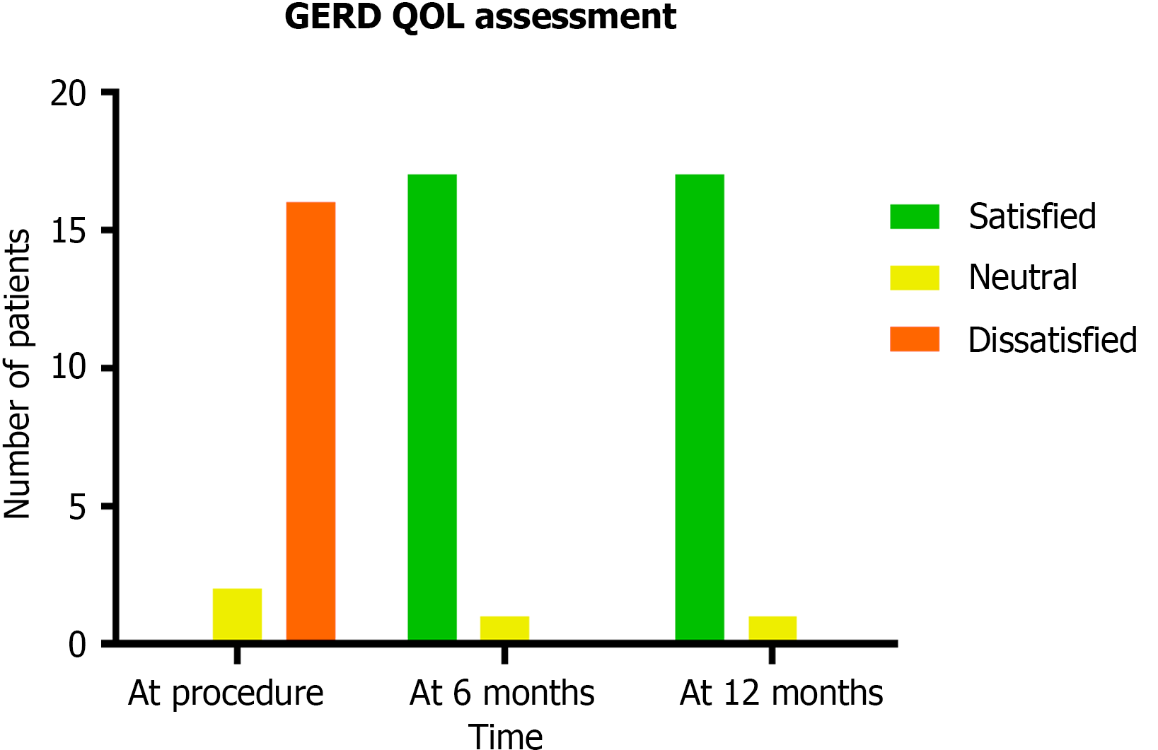
Figure 4 Gastroesophageal reflux disease -related quality of life at the time of the procedure and 6 and 12 months postoperatively (P < 0.
001). GERD: Gastroesophageal reflux disease; QOL: Quality of life.
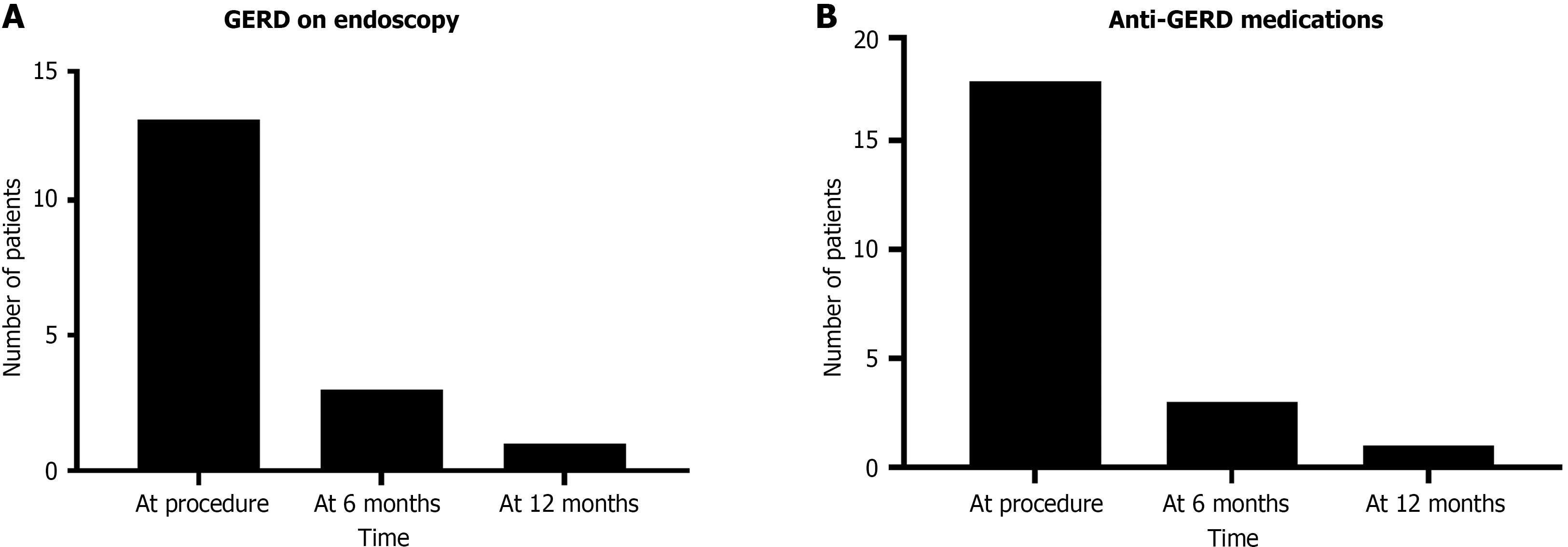
Figure 5 The detection of gastroesophageal reflux disease endoscopically and the use of anti-gastroesophageal reflux disease medications during the study period.
A: Decline in gastroesophageal reflux disease (GERD) detection endoscopically was statistically significant (P < 0.001); B: Decline in anti-GERD medication use was statistically significant (P < 0.001). GERD: Gastroesophageal reflux disease.
- Citation: Gadour E, Hoff AC. Gastric fundoplication with endoscopic technique: A novel approach for gastroesophageal reflux disease treatment. World J Gastrointest Endosc 2024; 16(10): 557-565
- URL: https://www.wjgnet.com/1948-5190/full/v16/i10/557.htm
- DOI: https://dx.doi.org/10.4253/wjge.v16.i10.557


