Copyright
©The Author(s) 2023.
World J Gastrointest Endosc. Sep 16, 2023; 15(9): 564-573
Published online Sep 16, 2023. doi: 10.4253/wjge.v15.i9.564
Published online Sep 16, 2023. doi: 10.4253/wjge.v15.i9.564
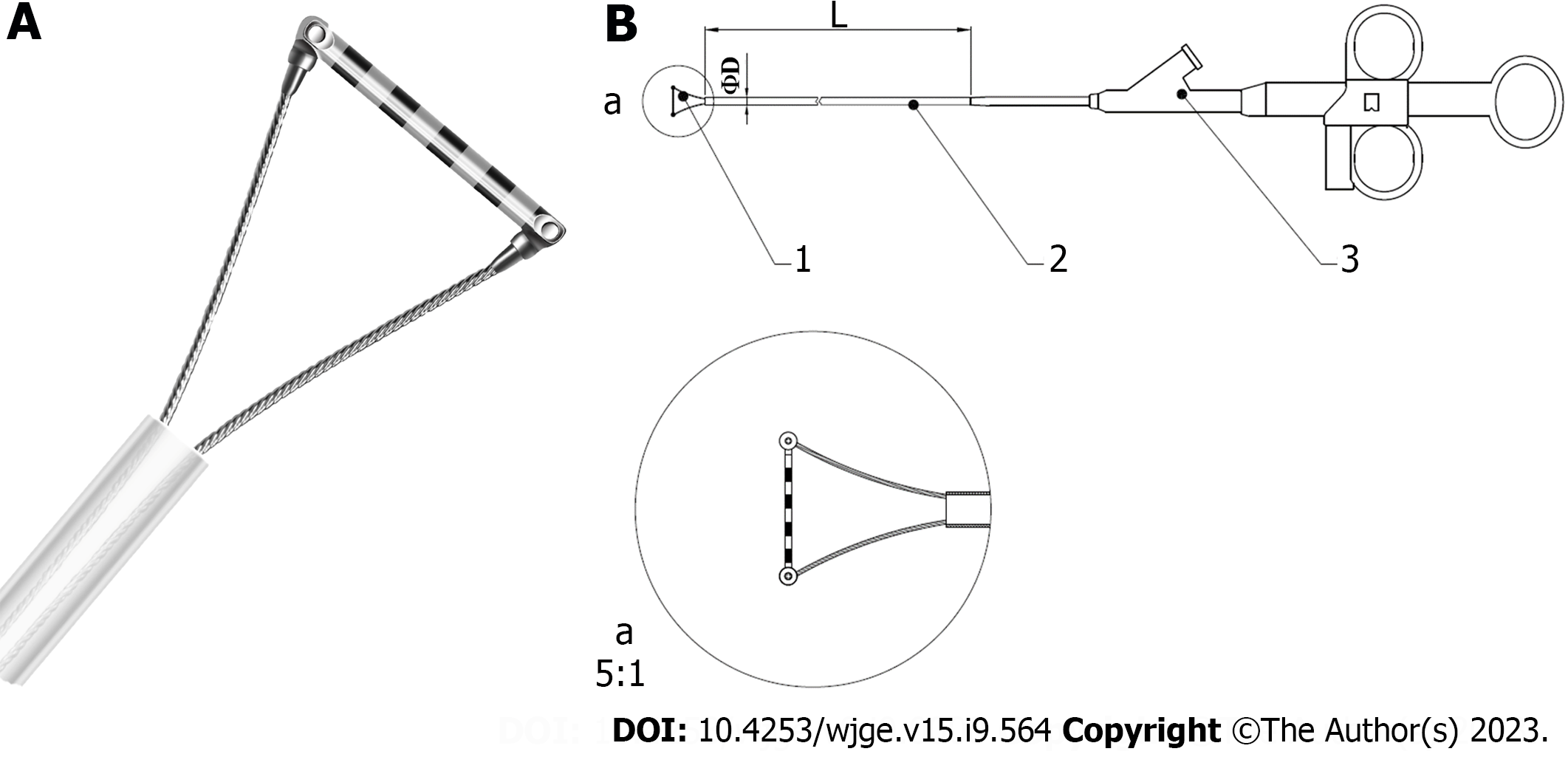
Figure 1 Diagram of Endoscopic Ruler.
A: The tip of Endoscopic Ruler; B: The structure of Endoscopic Ruler. 1: Tip; 2: Sheath; 3: Operating handle.
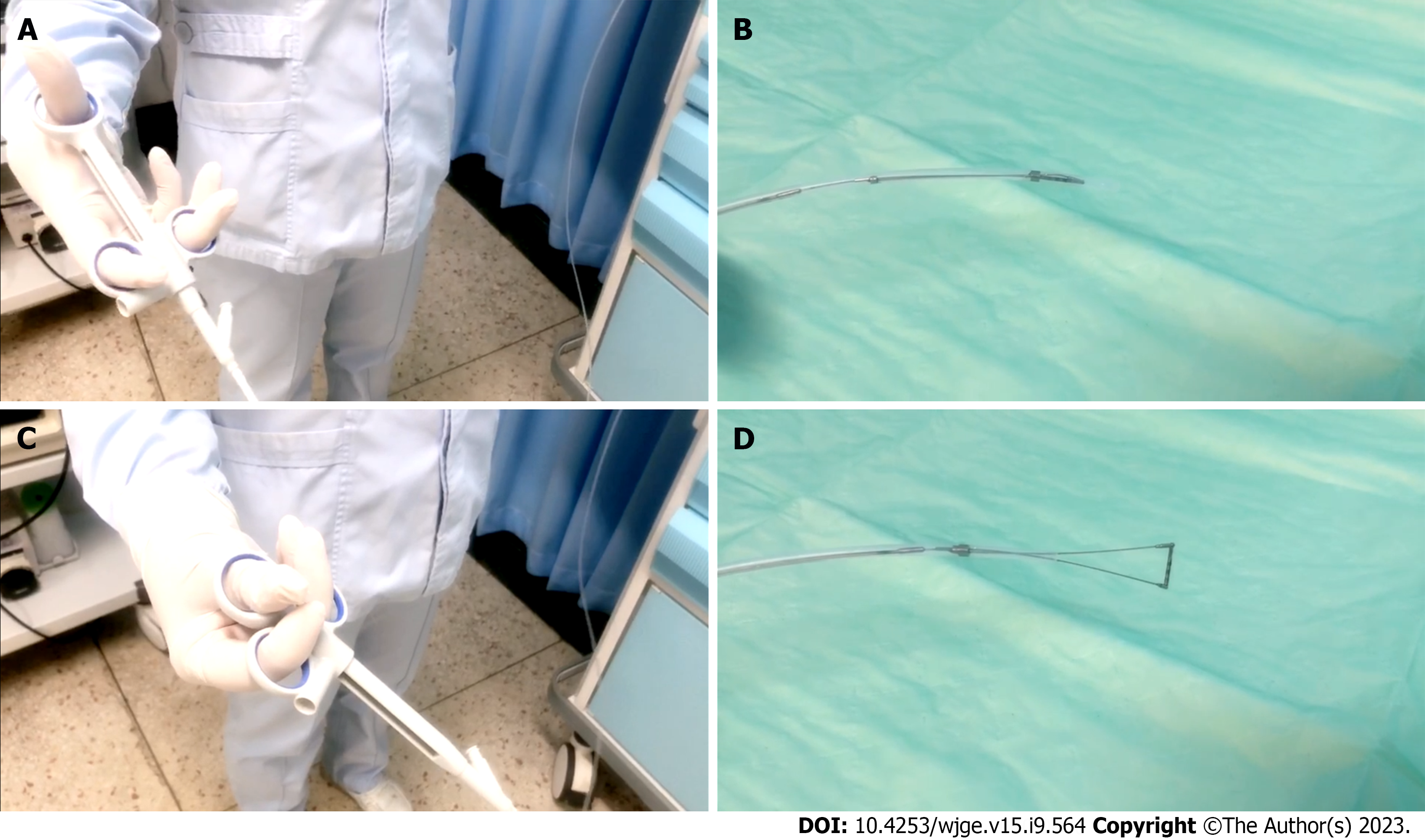
Figure 2 Measurement procedure of Endoscopic Ruler.
A and B: The operating handle of Endoscopic Ruler (A) when the tip is shut (B); C and D: The operating handle of Endoscopic Ruler (C) when the tip is open (D).
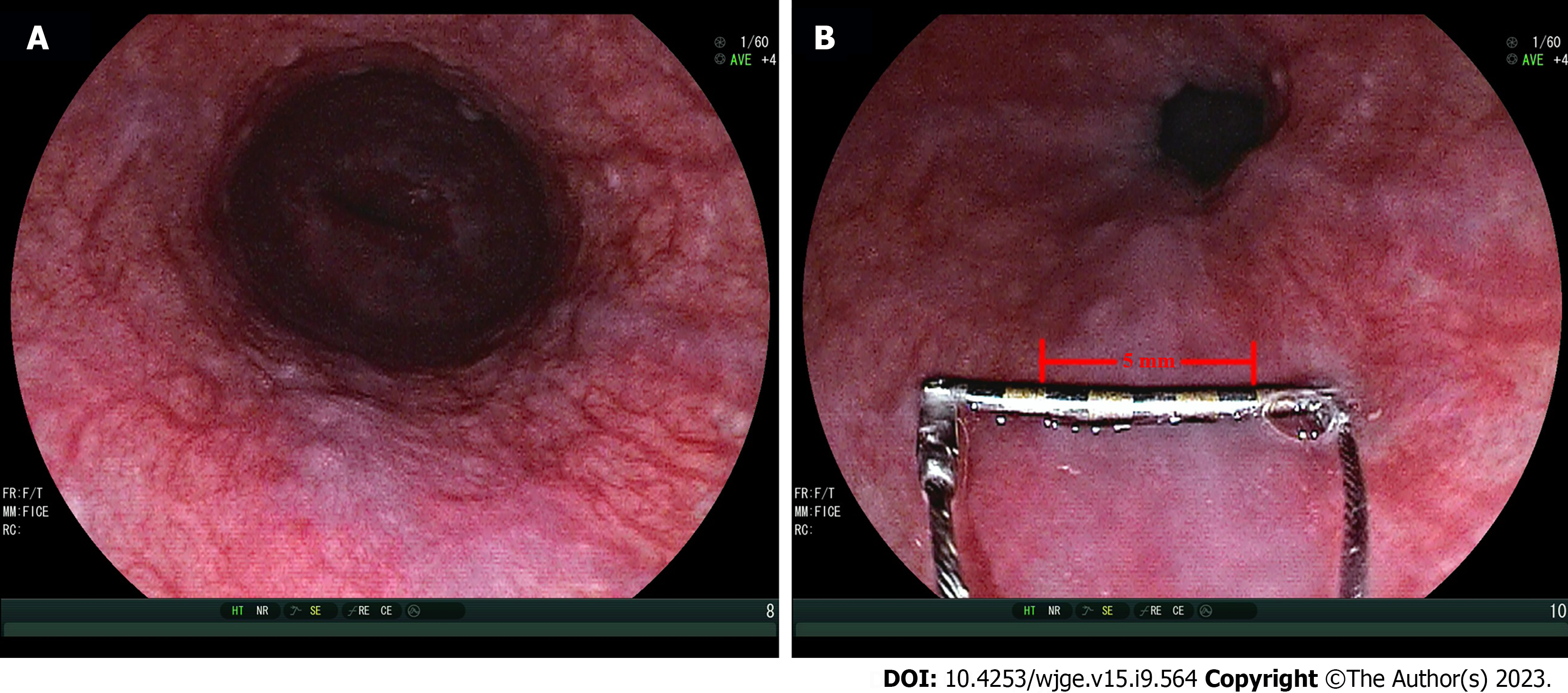
Figure 3 Representative examples of measurement of varix size by Endoscopic Ruler.
A: Small varices by endoscopists (varix size = 3-4 mm); B: Large varices by Endoscopic Ruler (varix size = 5 mm).
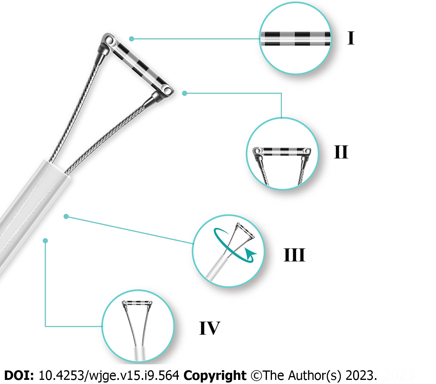
Figure 4 Diagram of improved Endoscopic Ruler.
I: Clear scale; II: Shorter tip; III: Synchronous rotation; IV: More smooth edge.
- Citation: Huang YF, Hu SJ, Bu Y, Li YL, Deng YH, Hu JP, Yang SQ, Shen Q, McAlindon M, Shi RC, Li XQ, Song TY, Qi HL, Jiao TW, Liu MY, He F, Zhu J, Ma B, Yu XB, Guo JY, Yu YH, Yong HJ, Yao WT, Ye T, Wang H, Dong WF, Liu JG, Wei Q, Tian J, Li XG, Dray X, Qi XL. Endoscopic Ruler for varix size measurement: A multicenter pilot study. World J Gastrointest Endosc 2023; 15(9): 564-573
- URL: https://www.wjgnet.com/1948-5190/full/v15/i9/564.htm
- DOI: https://dx.doi.org/10.4253/wjge.v15.i9.564


