Copyright
©The Author(s) 2023.
World J Gastrointest Endosc. May 16, 2023; 15(5): 386-396
Published online May 16, 2023. doi: 10.4253/wjge.v15.i5.386
Published online May 16, 2023. doi: 10.4253/wjge.v15.i5.386
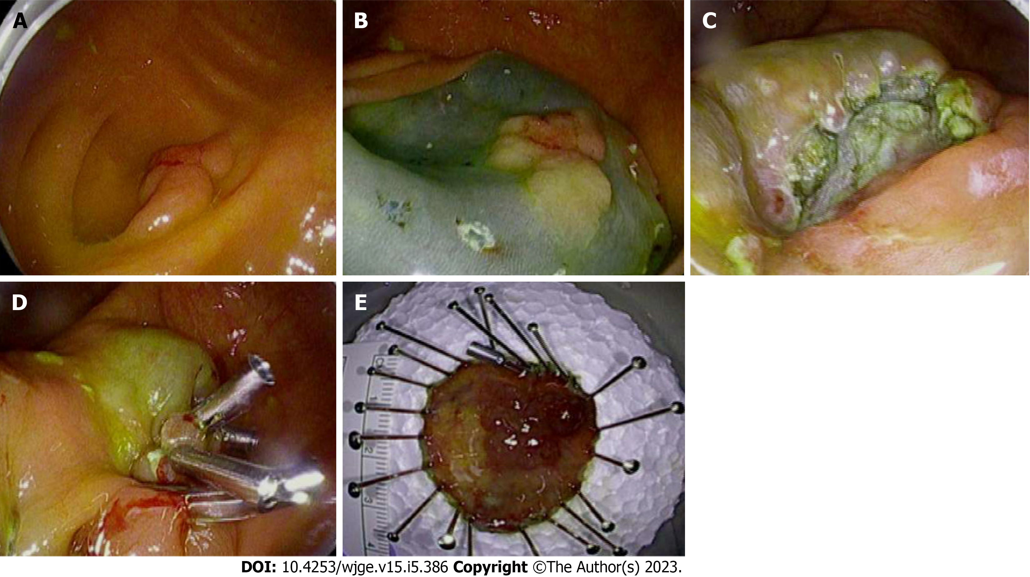
Figure 1 Step-by-step demonstration of a polyp removal via endoscopic submucosal dissection.
A: A 30 mm polyp occupying 50% of the appendiceal orifice circumference is visualized; B: The polyp borders are marked using the tip of the dual knife. Adequate lifting of the submucosa is achieved after the injection of Hespan Solution; C: The resection bed is seen after the dissection of the polyp from the underlying deeper layers; D: The defect is completely closed with 4 hemostatic clips; E: The result is an en bloc resection of the polyp.
- Citation: Patel AP, Khalaf MA, Riojas-Barrett M, Keihanian T, Othman MO. Expanding endoscopic boundaries: Endoscopic resection of large appendiceal orifice polyps with endoscopic mucosal resection and endoscopic submucosal dissection. World J Gastrointest Endosc 2023; 15(5): 386-396
- URL: https://www.wjgnet.com/1948-5190/full/v15/i5/386.htm
- DOI: https://dx.doi.org/10.4253/wjge.v15.i5.386


