Copyright
©The Author(s) 2023.
World J Gastrointest Endosc. May 16, 2023; 15(5): 368-375
Published online May 16, 2023. doi: 10.4253/wjge.v15.i5.368
Published online May 16, 2023. doi: 10.4253/wjge.v15.i5.368
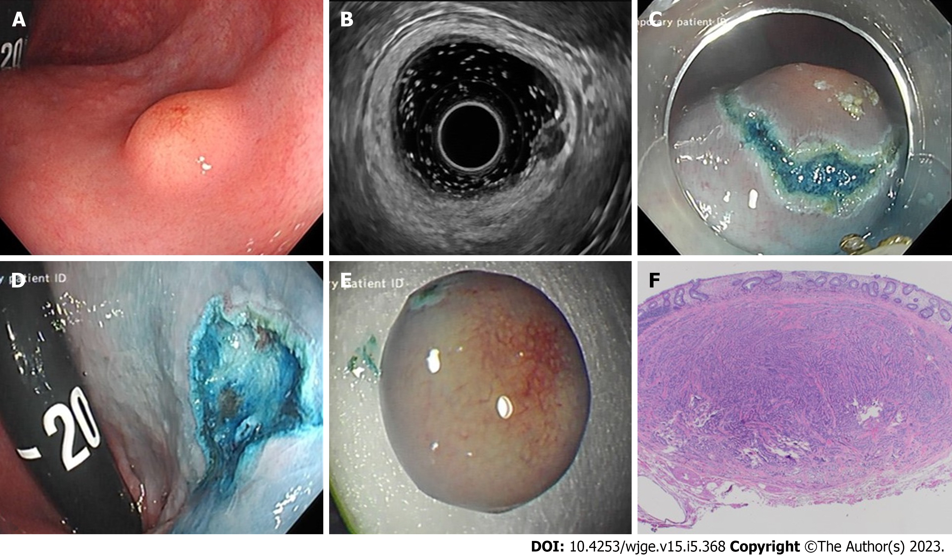
Figure 1 Case of a 33-year-old male with a background history of cystic fibrosis, referred for consideration for lung transplantation.
A: Endoscopic image of 6mm rectal neuroendocrine tumour (r-NRT) in retroflexion, 3 cm from anal verge; B: Endoscopic ultrasound images of the same 6 mm hypoechoic homogenous lesion, seen at 10 MHz frequency, consistent with a NET; C: Endoscopic image of hybrid Knife assisted snare resection approach; post circumferential submucosal incision; D: Endoscopic image of post en-bloc knife-assisted snare resection site in retroflexion; E: Excised en-bloc r-NET specimen; F: Neuroendocrine tumour composed of neuroendocrine cells arranged in anastomosing trabeculae with overlying rectal mucosa. The tumour is well circumscribed and has been excised (haematoxylin and eosin stain, 20× magnification).
- Citation: Keating E, Bennett G, Murray MA, Ryan S, Aird J, O'Connor DB, O'Toole D, Lahiff C. Rectal neuroendocrine tumours and the role of emerging endoscopic techniques. World J Gastrointest Endosc 2023; 15(5): 368-375
- URL: https://www.wjgnet.com/1948-5190/full/v15/i5/368.htm
- DOI: https://dx.doi.org/10.4253/wjge.v15.i5.368


