Copyright
©The Author(s) 2023.
World J Gastrointest Endosc. Apr 16, 2023; 15(4): 216-239
Published online Apr 16, 2023. doi: 10.4253/wjge.v15.i4.216
Published online Apr 16, 2023. doi: 10.4253/wjge.v15.i4.216
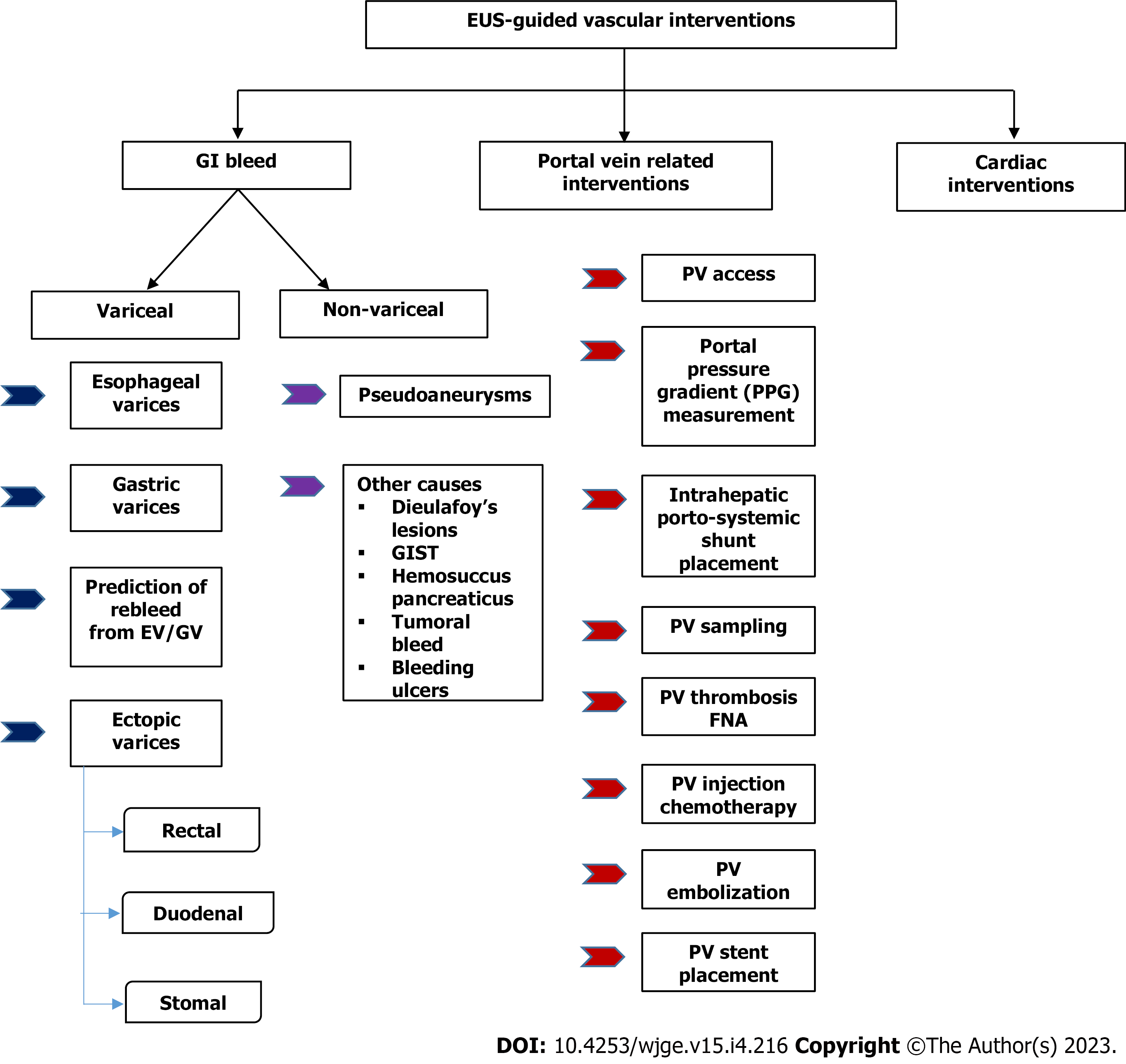
Figure 1 Flowchart of various endoscopic ultrasound guided vascular interventions.
EUS: Endoscopic ultrasound; GI: Gastrointestinal; EV: Esophageal varices; GV: Gastric varices; GIST: Gastrointestina stromal tumour; PV: Portal vein; FNA: Fine needle aspiration.
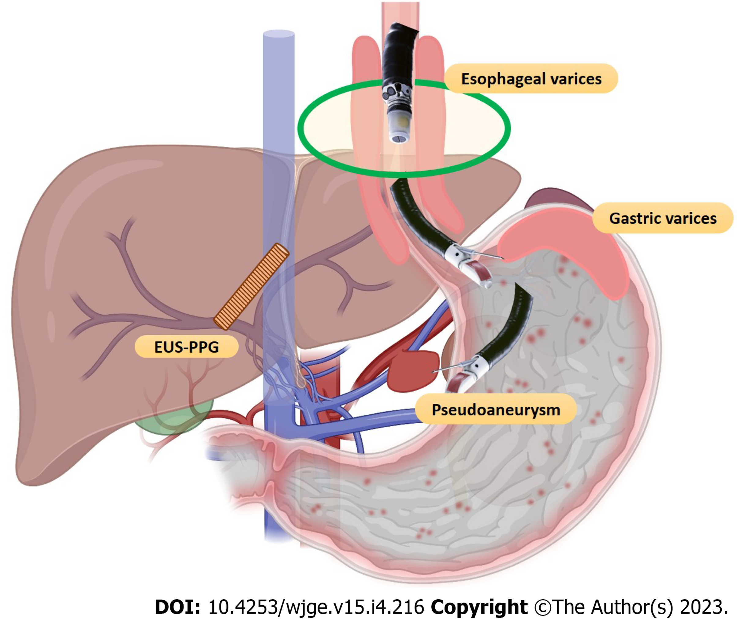
Figure 2 Spectrum of endoscopic ultrasound-guided vascular interventions.
EUS-PPG: Endoscopic ultrasound-portal pressure gradient.
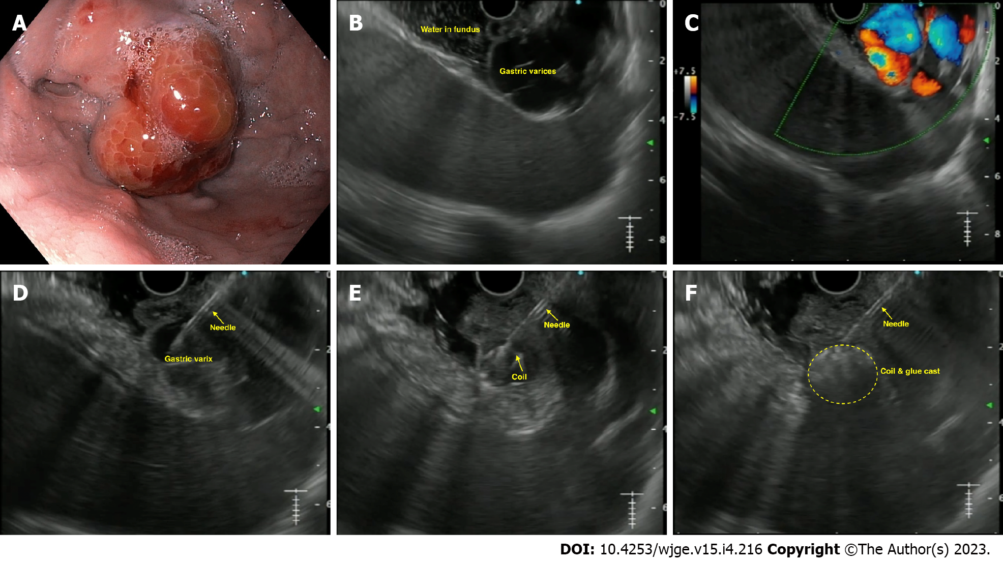
Figure 3 Endoscopic ultrasound-guided coil and glue injection for gastric varices.
A: Endoscopic image of gastric varix; B: Endoscopic ultrasound image of gastric varix; C: Colour Doppler showing flow in the varix; D: Puncture of the varix with 19-G needle; E: Coil being deployed in the varix; F: Glue injected leading to coil-glue cast with varix obliteration.
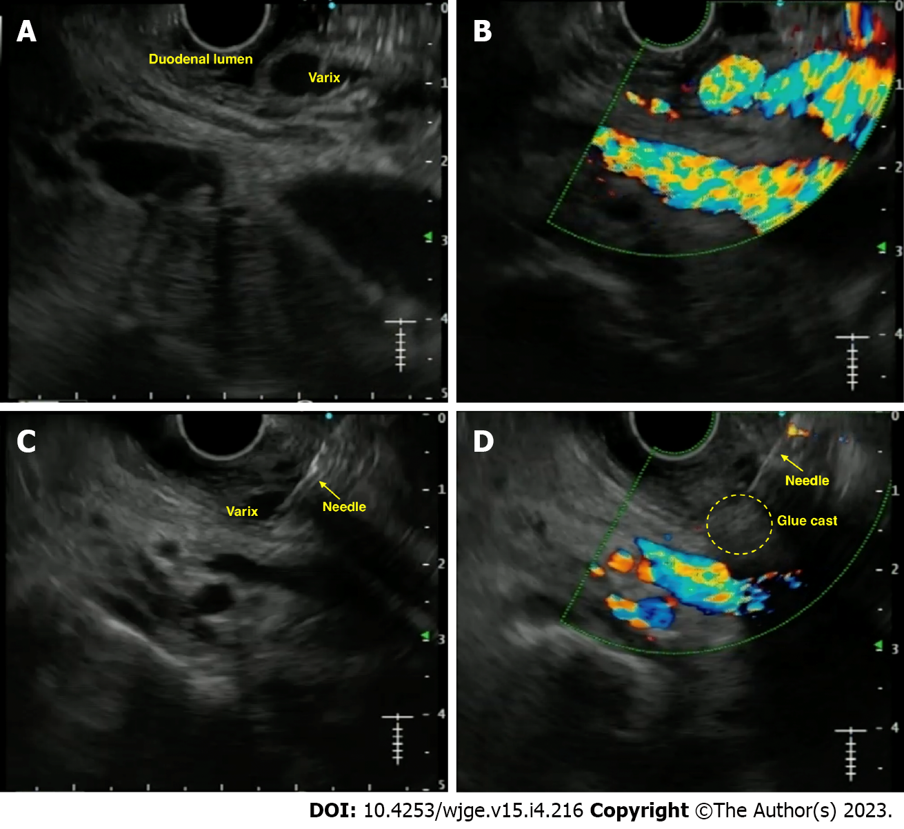
Figure 4 Endoscopic ultrasound-guided vascular therapy for duodenal varix.
A: Endoscopic ultrasound image of duodenal varix; B: Colour Doppler showing flow in the varix; C: Puncture of the varix with 19-G needle; D: Obliteration of the varix noted on Doppler flow.
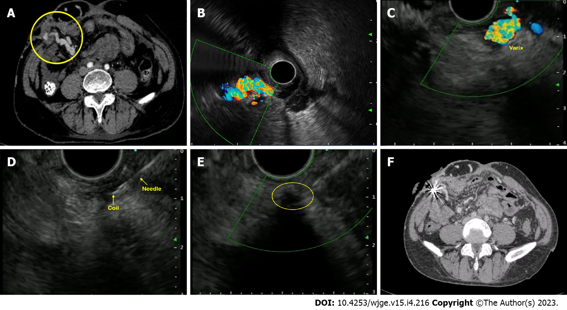
Figure 5 Endoscopic ultrasound-guided vascular therapy for parastomal varices.
A: Contrast enhanced computed tomography (CT) showing parastomal varices; B: Radial endoscopic ultrasound (EUS) image demonstrating the parastomal varices; C: Linear EUS image of the varices; D: Puncture of the varix with coil deployment; E: Obliteration of the varix with coil-glue cast; F: Post-intervention CT showing coil artifacts with obliteration of varices.
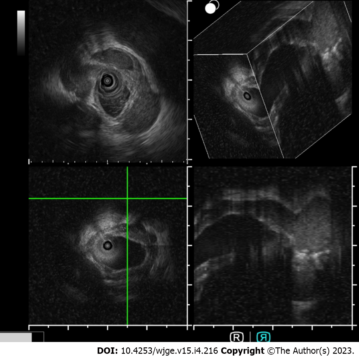
Figure 6 Intraductal ultrasound for pericholedochal varices.
Intraductal ultrasound using endoscopic ultrasound miniprobe (UM-DG20-31R IDUS probe, Olympus, Japan) for imaging in a case of portal cavernoma cholangiopathy with 3D reconstruction.
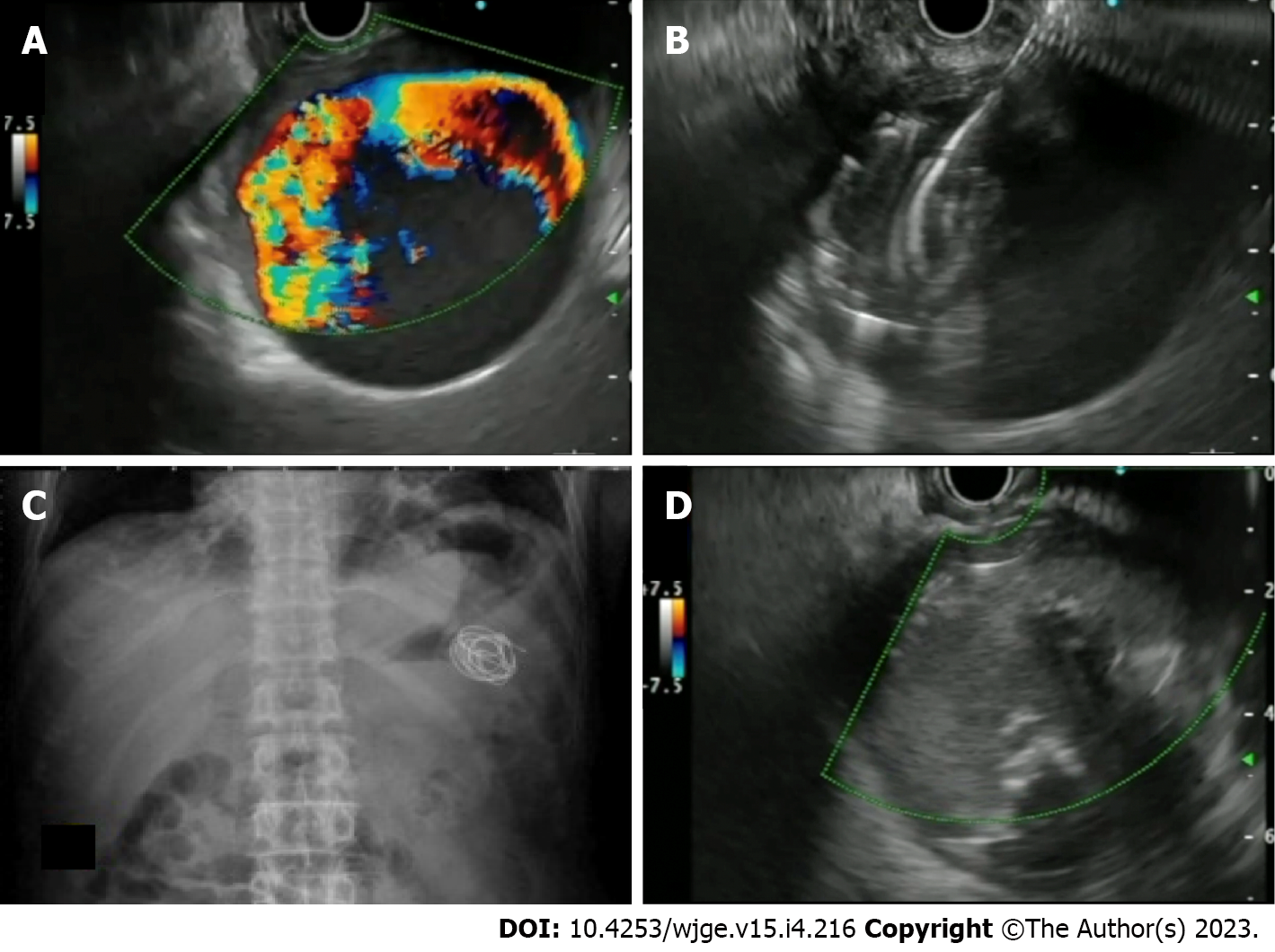
Figure 7 Endoscopic ultrasound-guided vascular therapy for pseudoaneurysm.
A: Giant splenic artery pesudoaneursym with Doppler flow; B: Puncture of the pseudoaneurysm with 19-G needle and deployment of coils; C: Abdominal X-ray showing deployed coils; D: Endoscopic ultrasound image of obliterated pseudoaneurysm after coil and glue injection.
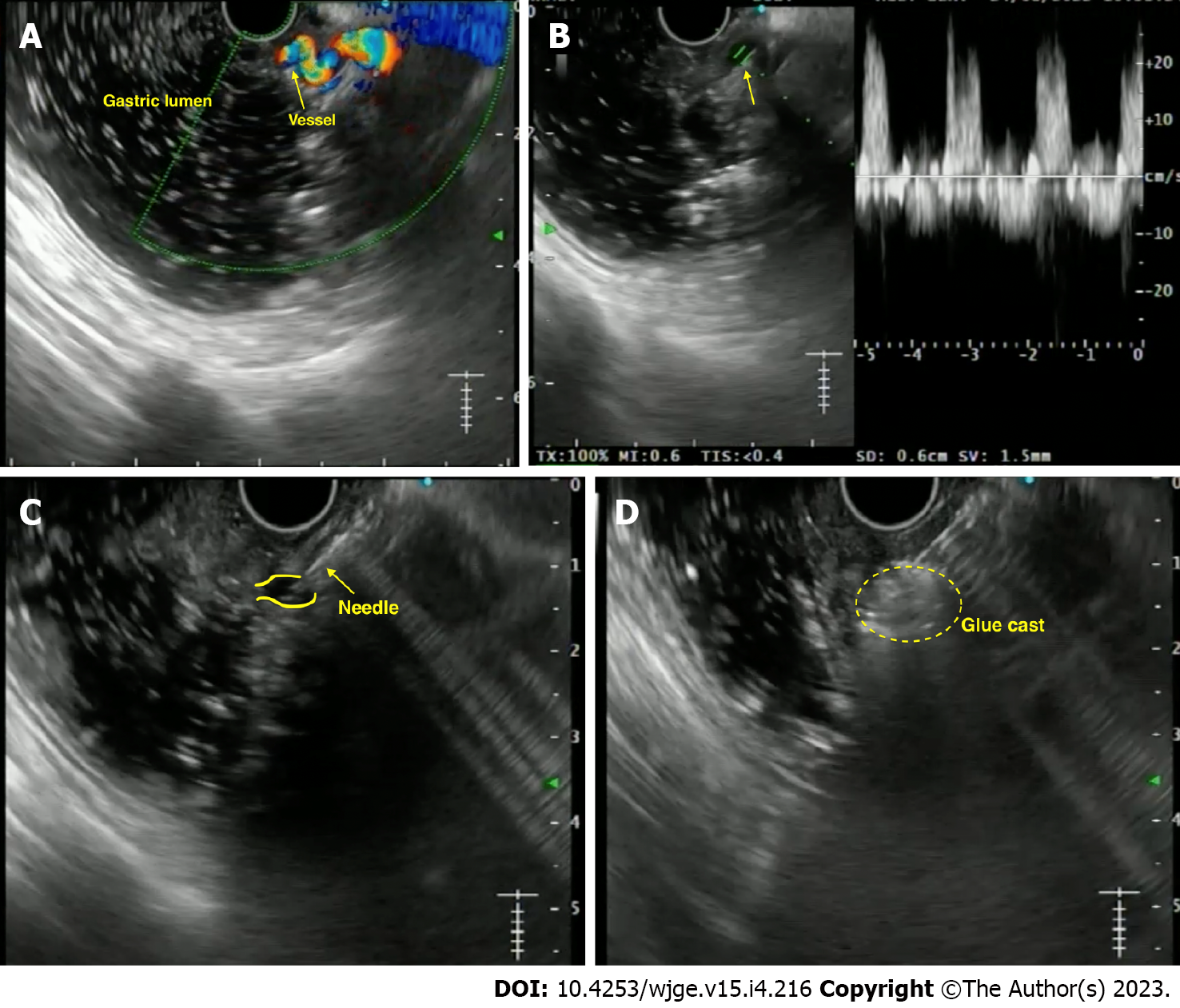
Figure 8 Endoscopic ultrasound-guided vascular therapy for dieulafoy’s lesion.
A: Endoscopic ultrasound image showing the culprit tortuous vessel coursing up to the mucosa; B: Power Doppler showing the flow pattern; C: Puncture of the vessel with a 22-G needle; D: Obliteration of the flow with formation of glue cast.
- Citation: Dhar J, Samanta J. Endoscopic ultrasound-guided vascular interventions: An expanding paradigm. World J Gastrointest Endosc 2023; 15(4): 216-239
- URL: https://www.wjgnet.com/1948-5190/full/v15/i4/216.htm
- DOI: https://dx.doi.org/10.4253/wjge.v15.i4.216


