Copyright
©The Author(s) 2023.
World J Gastrointest Endosc. Feb 16, 2023; 15(2): 32-43
Published online Feb 16, 2023. doi: 10.4253/wjge.v15.i2.32
Published online Feb 16, 2023. doi: 10.4253/wjge.v15.i2.32
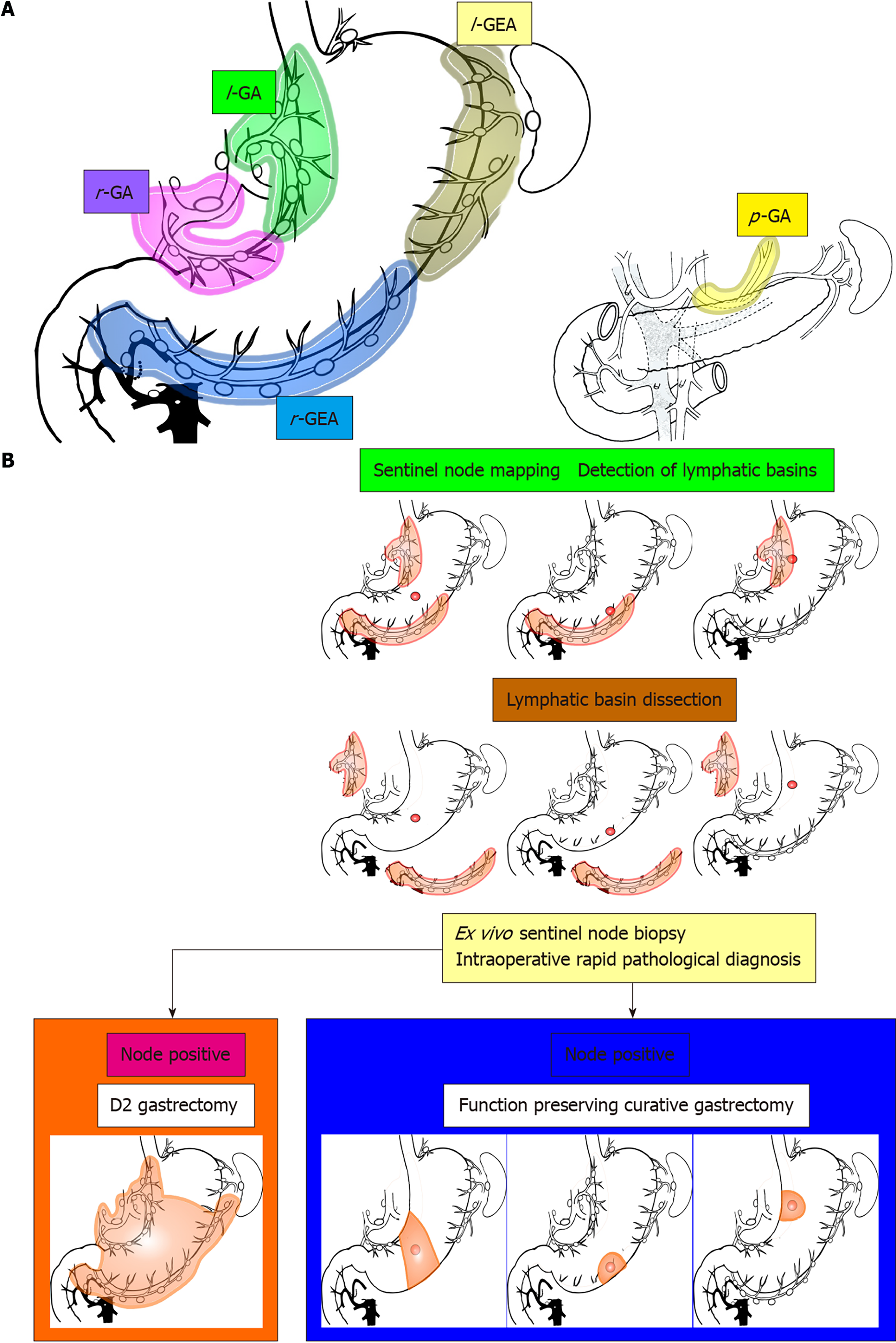
Figure 1 Lymphatic basins, lymphatic compartments, and the strategy of sentinel node navigation surgery.
l-GA: Left gastric artery basin; l-GEA: Left gastroepiploic artery basin; p-GA: Posterior gastric artery basin; r-GA: Right gastric artery basin; r-GEA: Right gastroepiploic artery basin. Citation: Kinami S, Nakamura N, Miyashita T, Kitakata H, Fushida S, Fujimura T, Iida Y, Inaki N, Ito T, Takamura H. Life prognosis of sentinel node navigation surgery for early-stage gastric cancer: Outcome of lymphatic basin dissection. World J Gastroenterol 2021; 27(46): 8010-8030. Copyright: The Authors 2021. Published by Baishideng Publishing Group Inc[38].
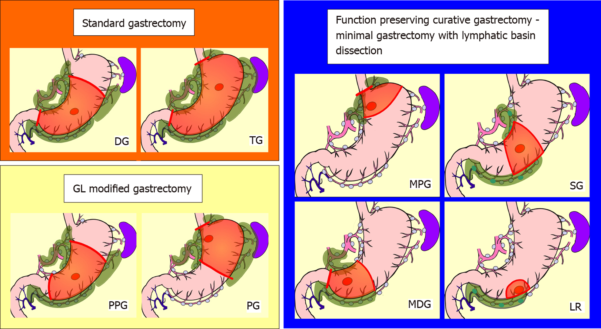
Figure 2 Schemas of standard gastrectomy, modified gastrectomy due to guidelines, and function-preserving curative gastrectomy with lymphatic basin dissection.
Red circle: tumor; Green-colored area: extent of lymph node dissection; Orange area: extent of gastrectomy. The extent of nodal dissection in standard gastrectomy and modified gastrectomy according to the guidelines was D1+. In contrast, the extent of nodal dissection in lymphatic basin dissection was defined as D0. DG: Distal gastrectomy; GL: Japanese gastric cancer treatment guidelines; LR: Local resection; MDG: Minidistal gastrectomy; MPG: Mini-proximal gastrectomy; PG: Proximal gastrectomy; PPG: Pylorus-preserving gastrectomy; SG: Segmental gastrectomy; TG: Total gastrectomy. Citation: Kinami S, Nakamura N, Miyashita T, Kitakata H, Fushida S, Fujimura T, Iida Y, Inaki N, Ito T, Takamura H. Life prognosis of sentinel node navigation surgery for early-stage gastric cancer: Outcome of lymphatic basin dissection. World J Gastroenterol 2021; 27(46): 8010-8030. Copyright: The Authors 2021. Published by Baishideng Publishing Group Inc[38].
Figure 3 Olympus Visera Elite II near-infrared fluorescence imaging system.
Copyright and courtesy of Olympus Europa SE & Co.KG, Hamburg, Germany.
Figure 4 Endoscopic submucosal indocyanine green injection in the stomach.
A: Pre-pyloric neoplastic lesion; B: Appearance after two submucosal indocyanine green (ICG) injections; C: Appearance after four circumferential submucosal ICG injections.
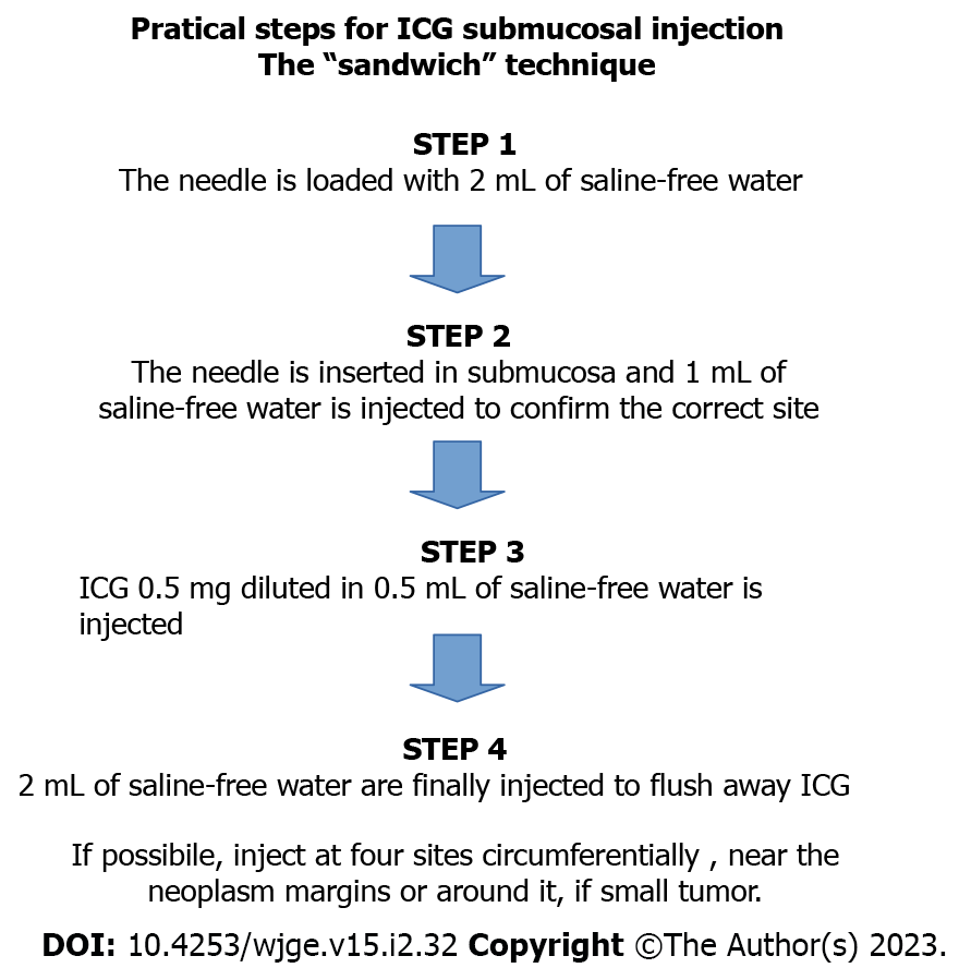
Figure 5 Practical steps for submucosal indocyanine green injection.
ICG: Indocyanine green.
- Citation: Calcara C, Cocciolillo S, Marten Canavesio Y, Adamo V, Carenzi S, Lucci DI, Premoli A. Endoscopic fluorescent lymphography for gastric cancer. World J Gastrointest Endosc 2023; 15(2): 32-43
- URL: https://www.wjgnet.com/1948-5190/full/v15/i2/32.htm
- DOI: https://dx.doi.org/10.4253/wjge.v15.i2.32


