Copyright
©The Author(s) 2023.
World J Gastrointest Endosc. Jan 16, 2023; 15(1): 10-18
Published online Jan 16, 2023. doi: 10.4253/wjge.v15.i1.10
Published online Jan 16, 2023. doi: 10.4253/wjge.v15.i1.10
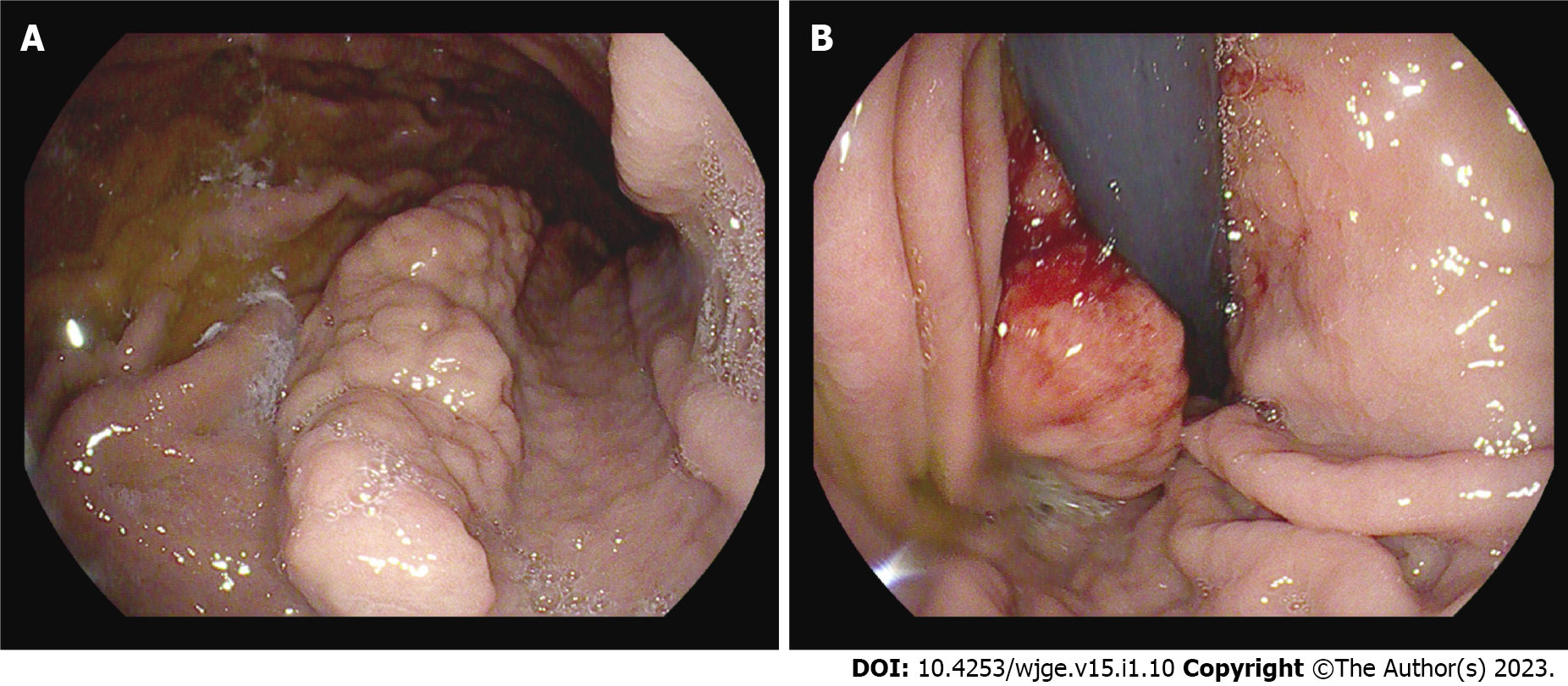
Figure 1 Polypoid mass found in the stomach during esophagogastroduodenoscopy.
A: A mass (4 cm × 1 cm) with enlarged mucosal folds in the body of the stomach between the lesser curvature and posterior wall; B: A small ulcer at the distal end of the mass.
Figure 2 Computed tomography of the abdomen with contrast enhancement.
A: A submucosal lesion in the stomach. Mean density of -89 HU suggested a submucosal lipoma; B: Length, 4.86 cm; C: Width, 1.96 cm.
Figure 3 Resection of the mass via tunneling endoscopic submucosal dissection.
A: A tunnel was created in the submucosal layer beneath the mass; B: Dissection was performed on both sides of the tunnel; C: Muscular defects were closed, and mucosal margins approximated with clips.
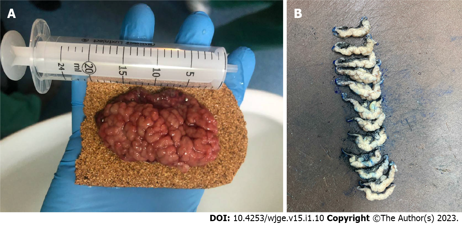
Figure 4 Resected specimen.
A: Immediately after resection, enlarged gastric folds were observed on the surface; B: Intersected specimen in the Pathology Department. Yellow tissue of the submucosal lipoma was observed.
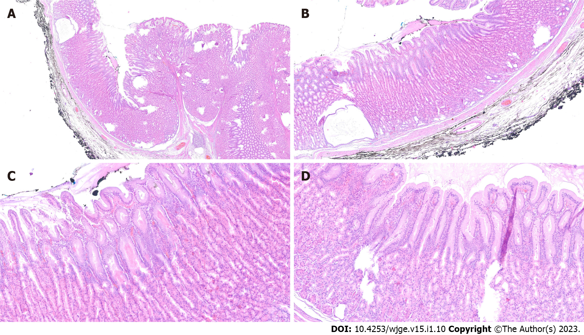
Figure 5 Histological changes in Ménétrier’s disease.
A: Low magnification; B: Cystic dilation of deep glands with foveolar hyperplasia; C: Foveolar hyperplasia with tortuous glands; D: Foveolar hyperplasia with dilation of the glands and oxyntic atrophy.
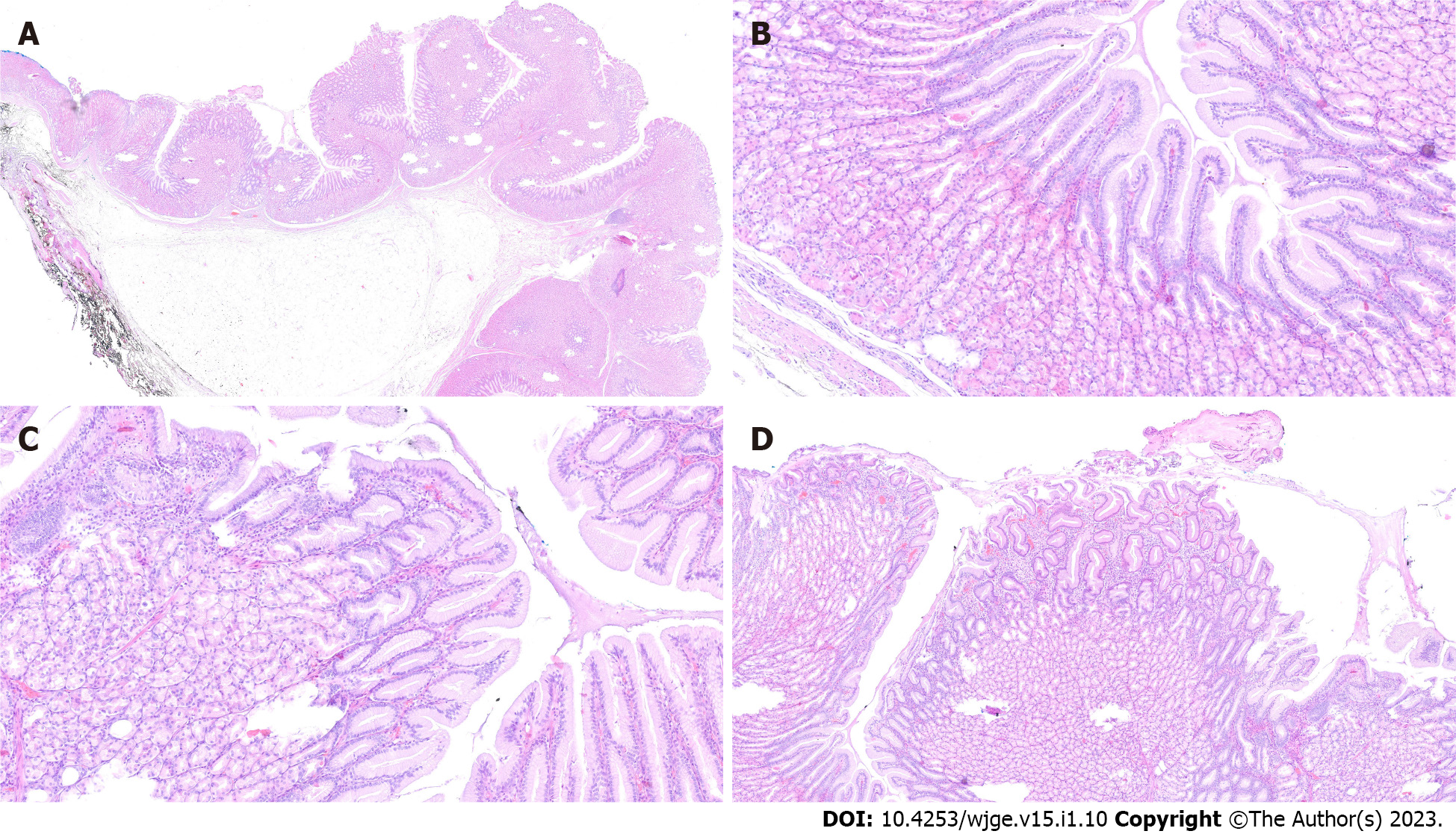
Figure 6 Submucosal lipoma and accompanying changes in the course of Ménétrier’s disease.
A: Low magnification; B: Foveolar hyperplasia; C: Foveolar hyperplasia with a corkscrew morphology; D: Foveolar hyperplasia with tortuous glands and mild inflammation in lamina propria.
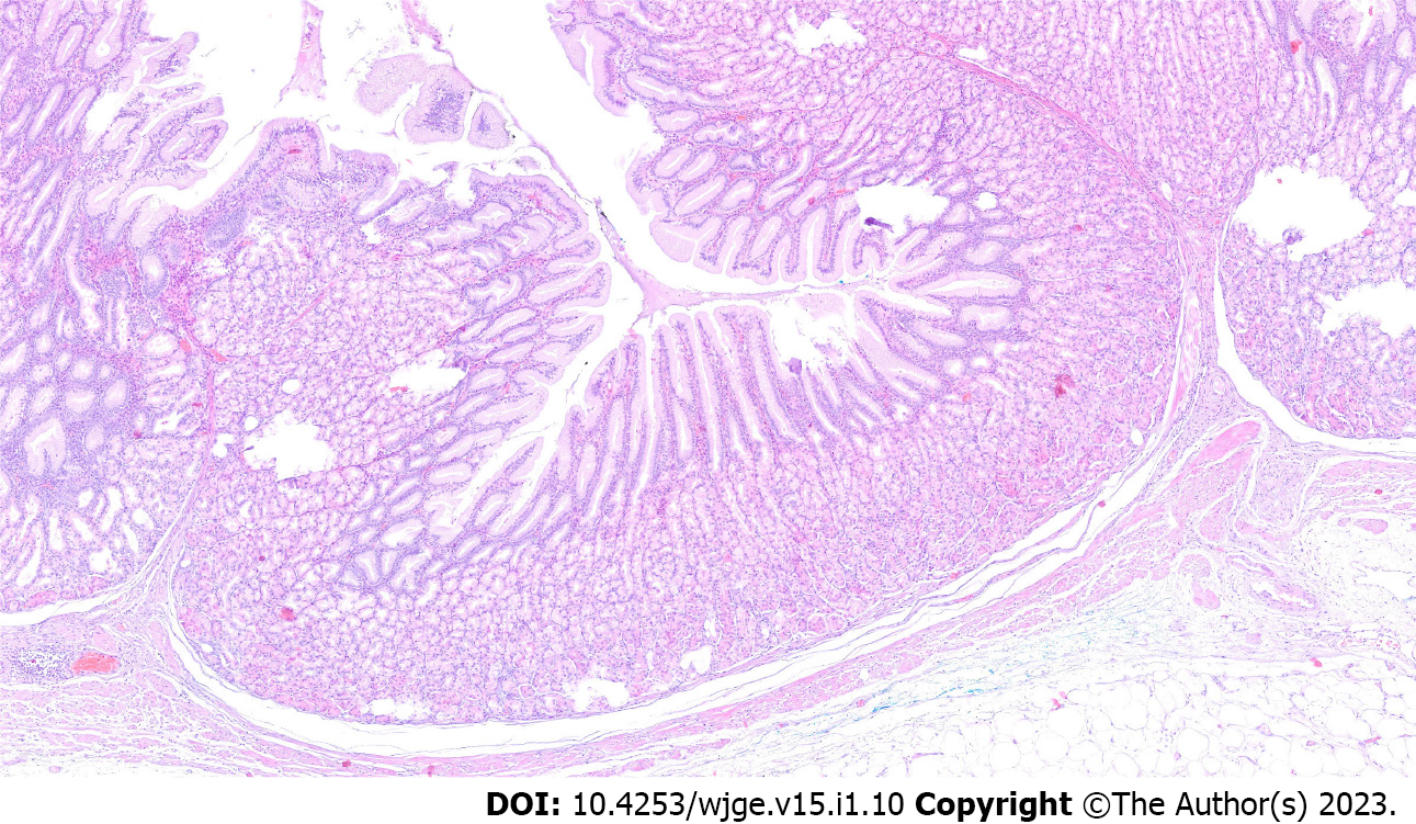
Figure 7 Histological changes in the course of Ménétrier’s disease: Foveolar hyperplasia, proliferation of muscularis mucosae and mild inflammation of lamina propria.
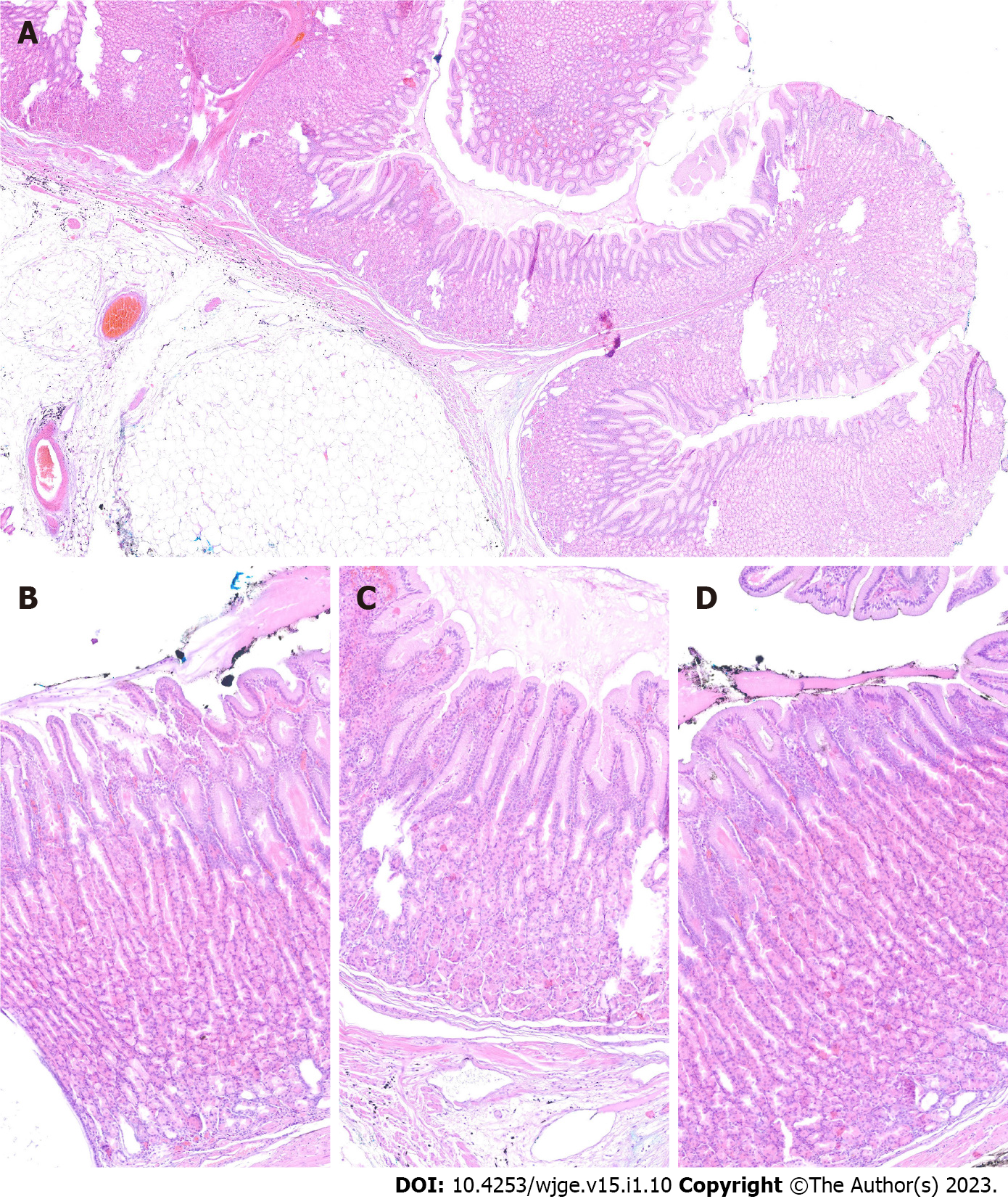
Figure 8 The mucosa with changes in the course of Ménétrier’s disease and the adjacent submucosal lipoma.
The lipoma was adjacent to the mucosa without crossing its borders. A: Low magnification; B, C and D: Representative images taken at high magnification.
- Citation: Kmiecik M, Walczak A, Samborski P, Paszkowski J, Dobrowolska A, Karczewski J, Swora-Cwynar E. Upper gastrointestinal bleeding as an unusual manifestation of localized Ménétrier’s disease with an underlying lipoma: A case report. World J Gastrointest Endosc 2023; 15(1): 10-18
- URL: https://www.wjgnet.com/1948-5190/full/v15/i1/10.htm
- DOI: https://dx.doi.org/10.4253/wjge.v15.i1.10


