Copyright
©The Author(s) 2022.
World J Gastrointest Endosc. Sep 16, 2022; 14(9): 564-574
Published online Sep 16, 2022. doi: 10.4253/wjge.v14.i9.564
Published online Sep 16, 2022. doi: 10.4253/wjge.v14.i9.564
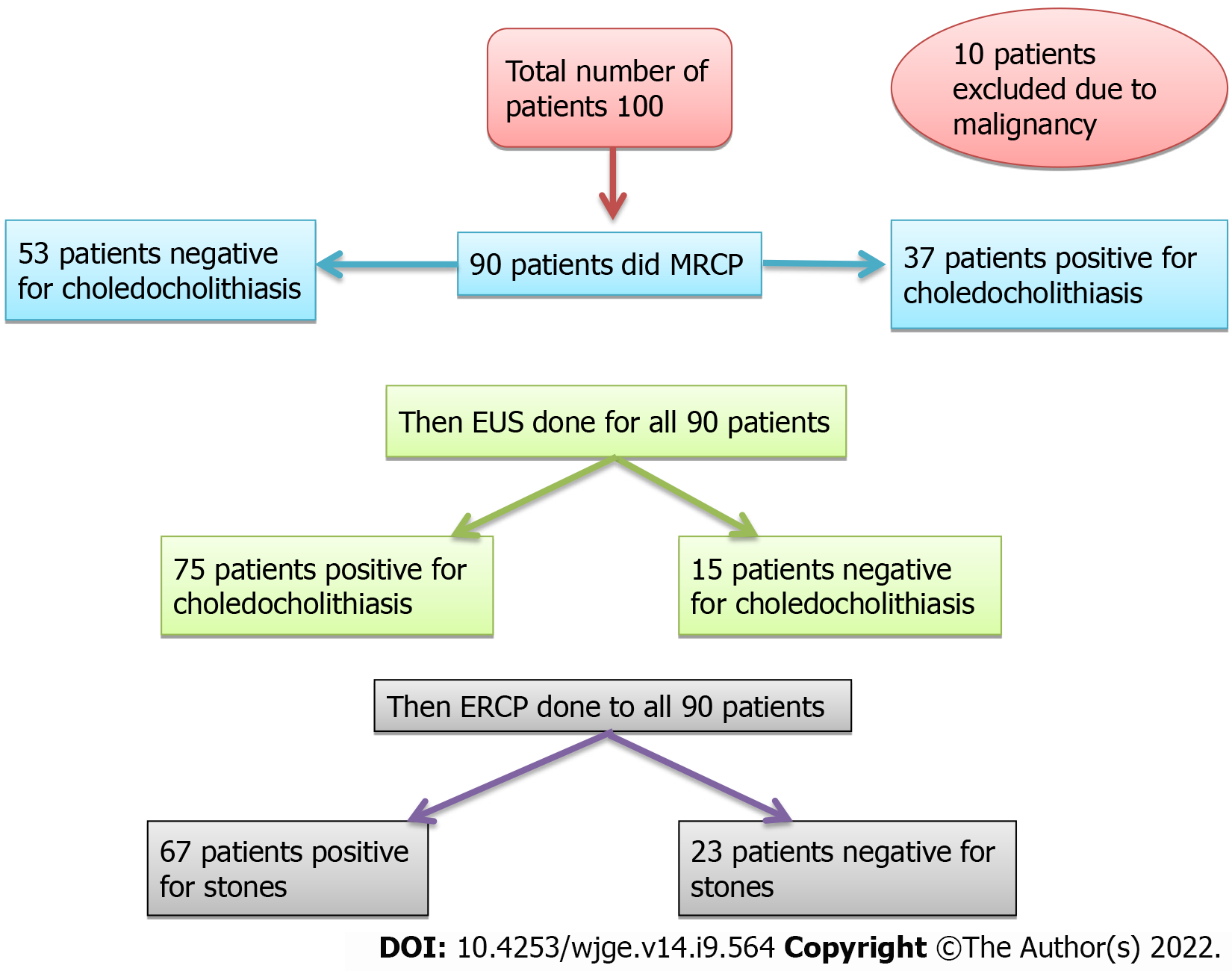
Figure 1 Flow chart of the studied patients.
MRCP: Magnetic resonance cholangiopancreatography: EUS: Endoscopic ultrasound; ERCP: Endoscopic Retrograde Cholangiopancreatography.
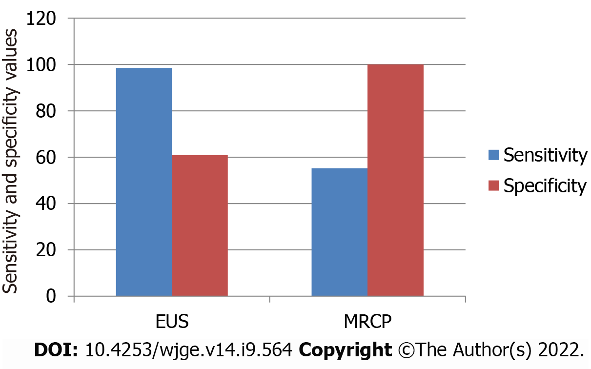
Figure 2 Comparison of sensitivity and specificity of endoscopic ultrasound and magnetic resonance cholangiopancreatography in detecting choledocholithiasis.
EUS: Endoscopic ultrasound; MRCP: Magnetic resonance cholangiopancreatography.
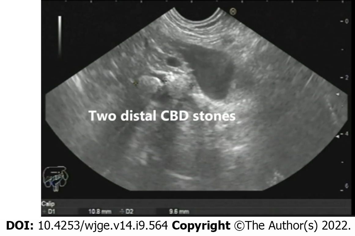
Figure 3 Two distal common bile duct stones as seen from the gastric body.
CBD: Common bile duct.
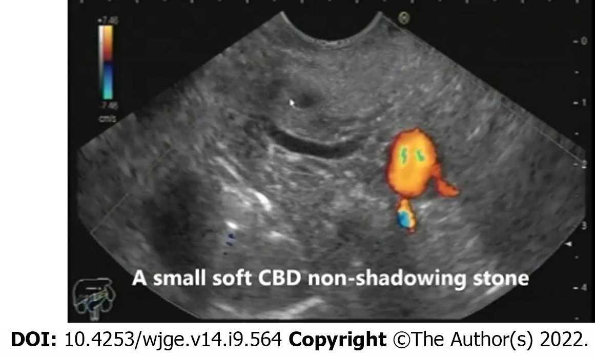
Figure 4 A small soft non-shadowing common bile duct stone as seen from the bulb of the duodenum.
CBD: Common bile duct.
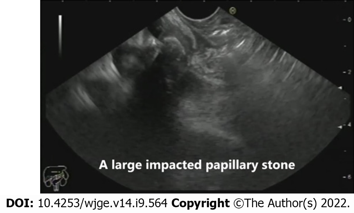
Figure 5 An impacted stone in the region of the major papilla as seen in the mid-second part of the duodenum.
- Citation: Eissa M, Okasha HH, Abbasy M, Khamis AK, Abdellatef A, Rady MA. Role of endoscopic ultrasound in evaluation of patients with missed common bile duct stones. World J Gastrointest Endosc 2022; 14(9): 564-574
- URL: https://www.wjgnet.com/1948-5190/full/v14/i9/564.htm
- DOI: https://dx.doi.org/10.4253/wjge.v14.i9.564


