Copyright
©The Author(s) 2022.
World J Gastrointest Endosc. May 16, 2022; 14(5): 342-350
Published online May 16, 2022. doi: 10.4253/wjge.v14.i5.342
Published online May 16, 2022. doi: 10.4253/wjge.v14.i5.342
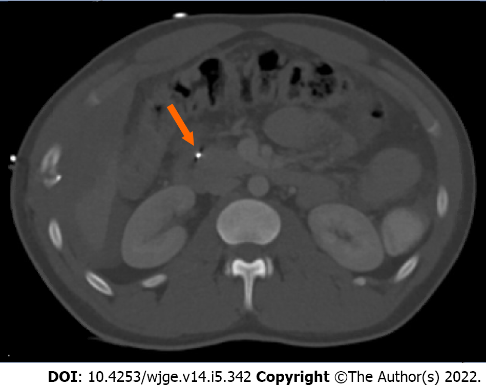
Figure 1 Computed tomography of the abdomen demonstrating bullet shrapnel involving the proximal duodenum and the pancreatic head (arrow).
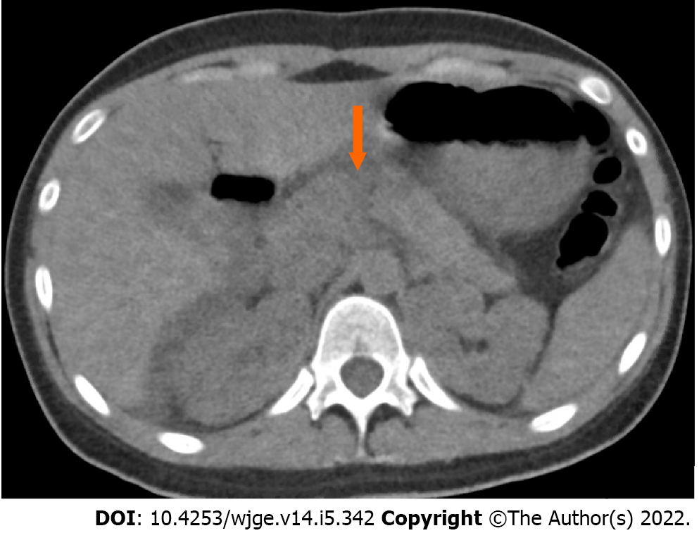
Figure 2 Computed tomography of the abdomen revealing a full-thickness pancreatic transection involving the proximal tail and neck (arrow).
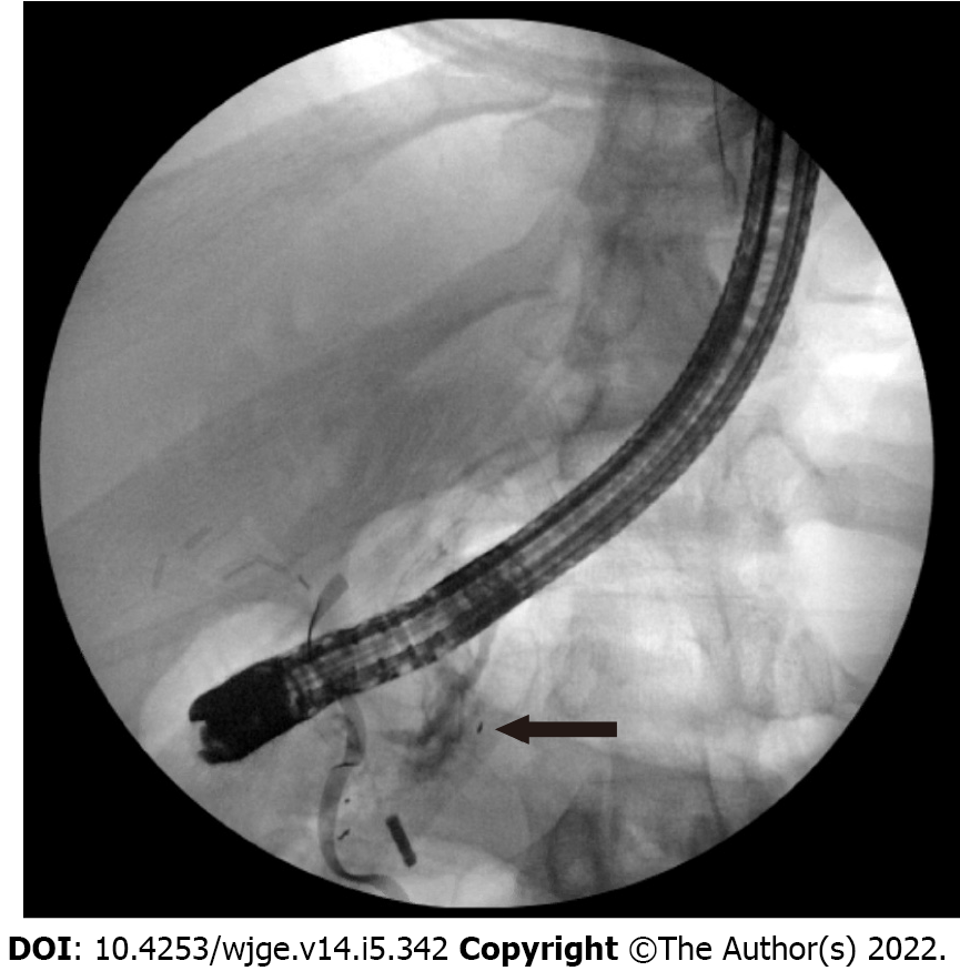
Figure 3 Endoscopic retrograde cholangiopancreatography fluoroscopy showing a ventral pancreatic ductal leak in the head of the pancreas (arrow).
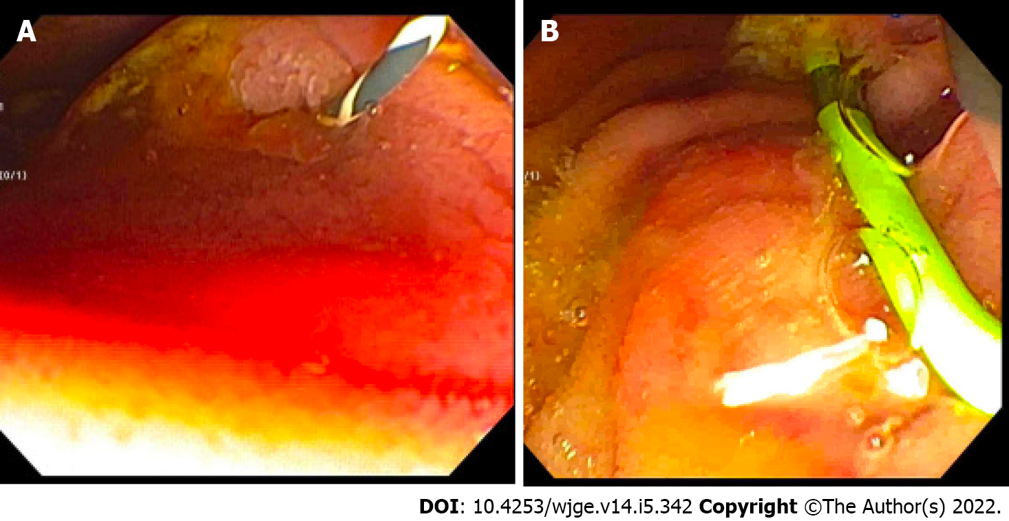
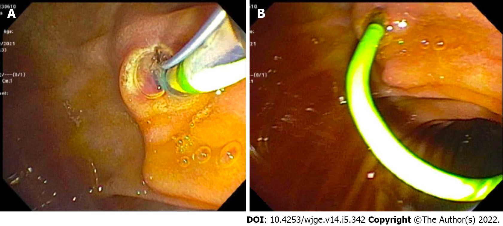
Figure 4 Intraoperative endoscopic retrograde cholangiopancreatography.
A and B: Endoscopic view following placement of an angled Visiglide wire into the ventral pancreatic duct (A) and placement of a plastic stent in the dorsal pancreatic duct (B).
Figure 5 Endoscopic view of the pancreatic sphincterotomy and pancreatic duct plastic stent placement.
A: Pancreatic sphincterotomy; B: Pancreatic duct plastic stent placement.
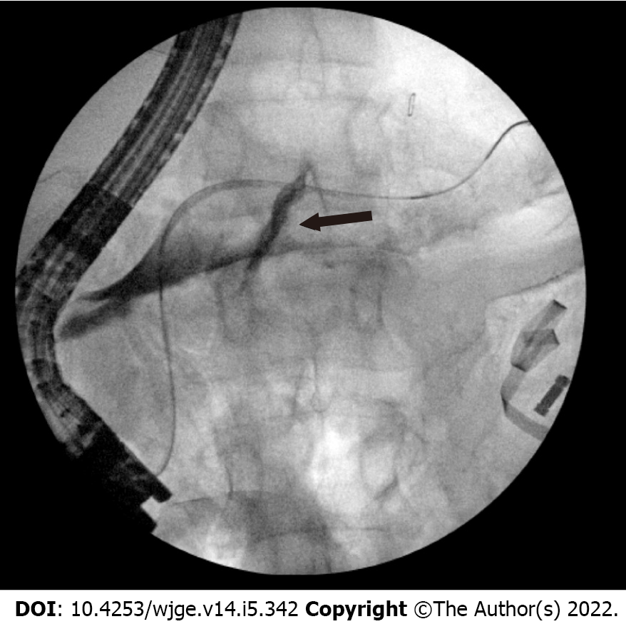
Figure 6 Endoscopic retrograde cholangiopancreatography fluoroscopic view demonstrating a dorsal pancreatic ductal leak (arrow).
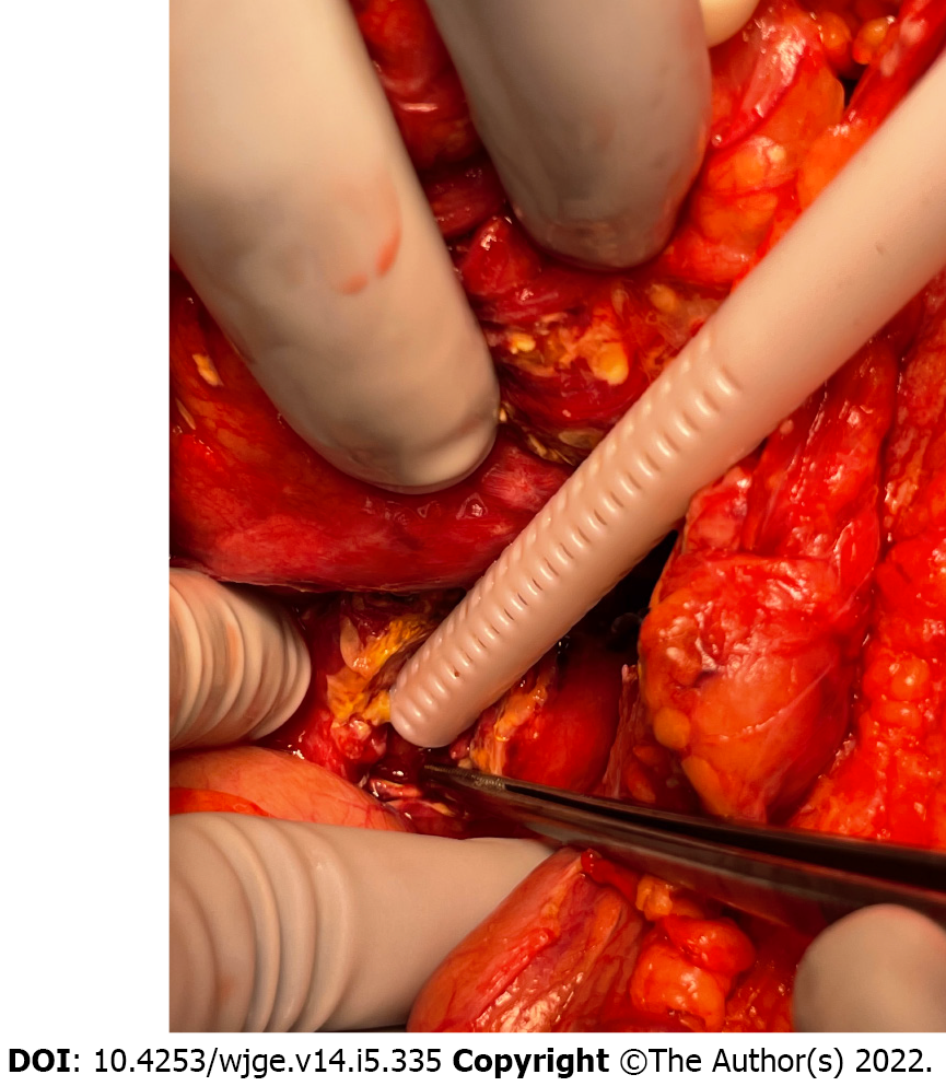
Figure 7 Intraoperative photo confirming following placement of the pancreatic ductal stent.
- Citation: Canakis A, Kesar V, Hudspath C, Kim RE, Scalea TM, Darwin P. Intraoperative endoscopic retrograde cholangiopancreatography for traumatic pancreatic ductal injuries: Two case reports. World J Gastrointest Endosc 2022; 14(5): 342-350
- URL: https://www.wjgnet.com/1948-5190/full/v14/i5/342.htm
- DOI: https://dx.doi.org/10.4253/wjge.v14.i5.342


