Copyright
©The Author(s) 2022.
World J Gastrointest Endosc. May 16, 2022; 14(5): 335-341
Published online May 16, 2022. doi: 10.4253/wjge.v14.i5.335
Published online May 16, 2022. doi: 10.4253/wjge.v14.i5.335
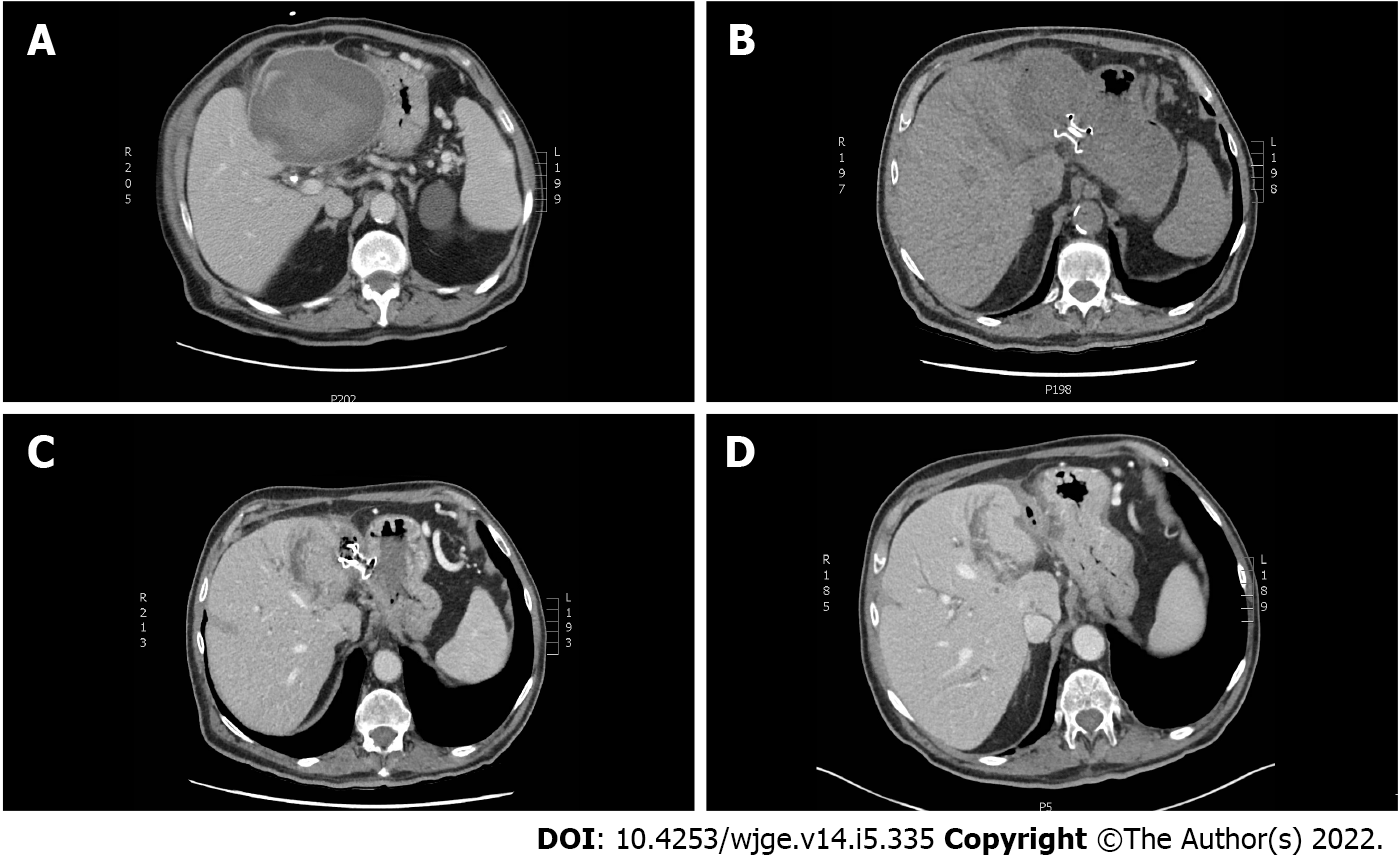
Figure 1 Computed tomography scan of the upper abdomen showing the left subcapsular hepatic hematoma at different stages of endoscopic treatment.
A: At diagnosis, the subcapsular hepatic hematoma (SHH) size was 12.5 cm × 10.5 cm × 12.5 cm and was compressing the stomach; B: At day 1 after endoscopic ultrasonography and lumen apposing metal stent (LAMS) was installed by transgastric approach; C: A month later, a control computed tomography (CT) scan of the upper abdomen showed a resorption of the SHH after 4 debridement sessions; D: Control CT scan of the upper abdomen after the LAMS was removed endoscopically.
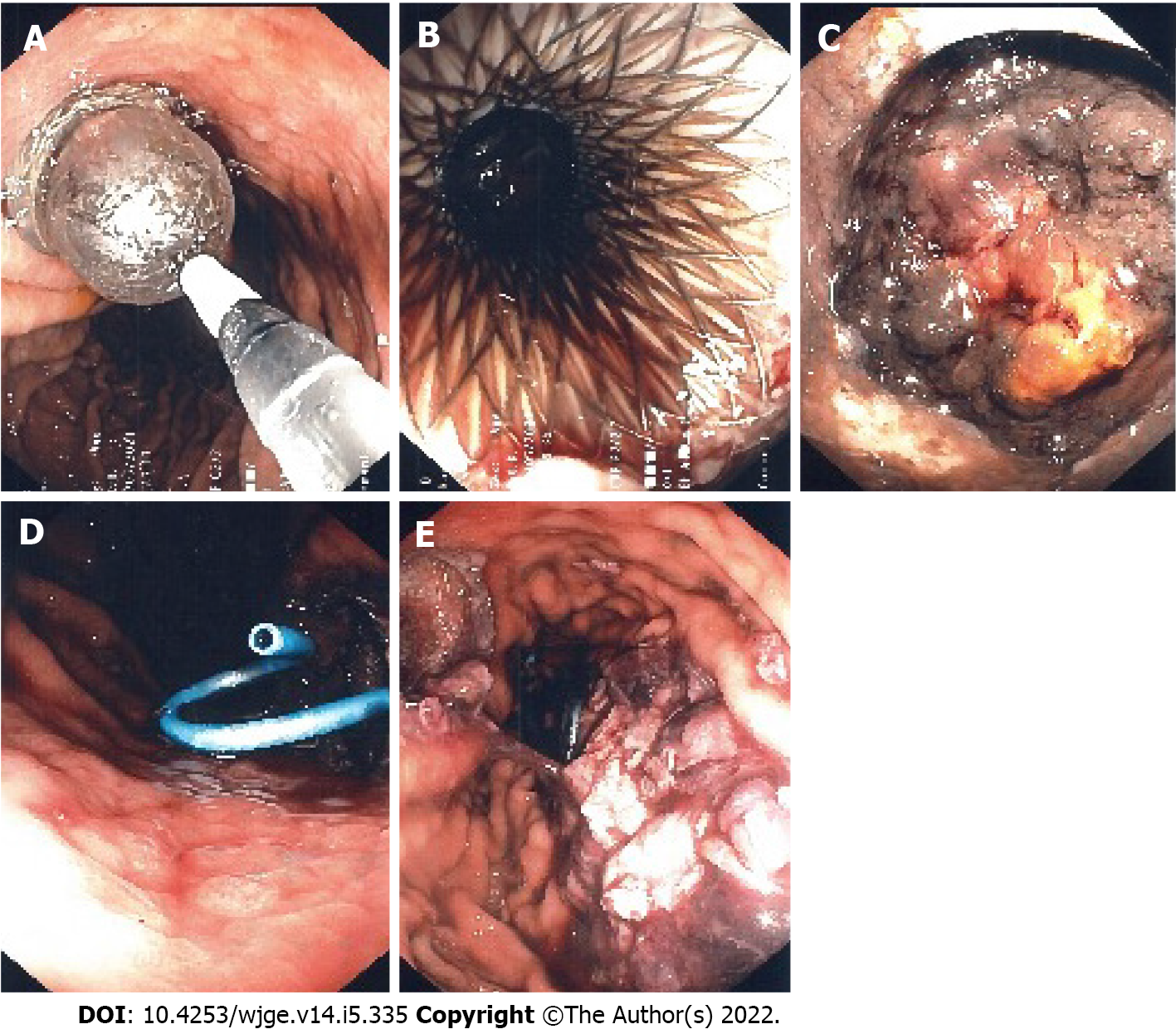
Figure 2 Endoscopic ultrasonography guided transgastric insertion of fully covered 10 mm × 15 mm lumen apposing metal stent allows endoscopic access to the subcapsular hepatic hematoma for drainage and debridement.
A: Dilatation of the lumen apposing metal stent (LAMS) was needed for the first debridement; B: Endoscopic image showing the LAMS after dilatation during the first of four debridements; C: Endoscopic image showing the subcapsular hepatic hematoma (SHH) during the second debridement; D: After each debridement, a double pigtail stent was inserted into the lumen of the LAMS allowing a more complete drainage of the SHH; E: Endoscopic image showing debris of the hematoma inside the stomach after the last debridement.
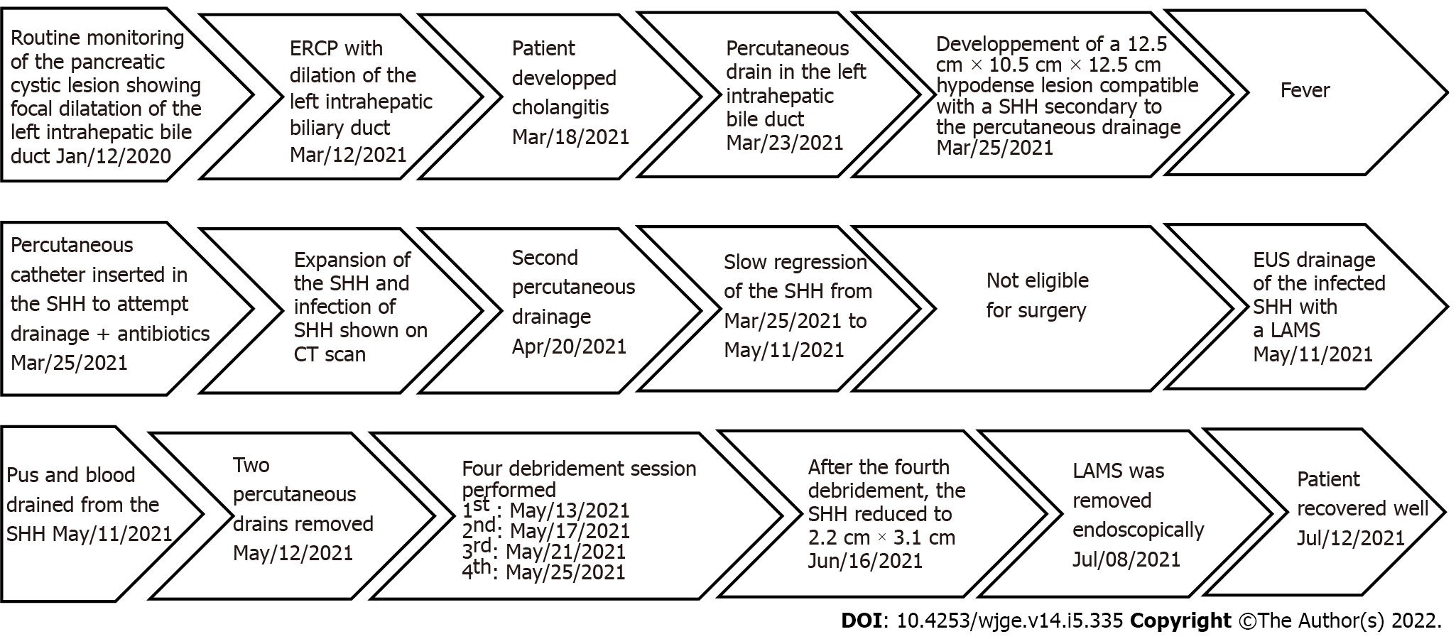
Figure 3 Timeline of the medical care episode.
CT: Computed tomography; SHH: Subcapsular hepatic hematoma; ERCP: Endoscopic retrograde cholangiopancreatography; EUS: Endoscopic ultrasonography; LAMS: Lumen apposing metal stent.
- Citation: Doyon T, Maniere T, Désilets É. Endoscopic ultrasonography drainage and debridement of an infected subcapsular hepatic hematoma: A case report. World J Gastrointest Endosc 2022; 14(5): 335-341
- URL: https://www.wjgnet.com/1948-5190/full/v14/i5/335.htm
- DOI: https://dx.doi.org/10.4253/wjge.v14.i5.335


