Copyright
©The Author(s) 2022.
World J Gastrointest Endosc. May 16, 2022; 14(5): 250-266
Published online May 16, 2022. doi: 10.4253/wjge.v14.i5.250
Published online May 16, 2022. doi: 10.4253/wjge.v14.i5.250
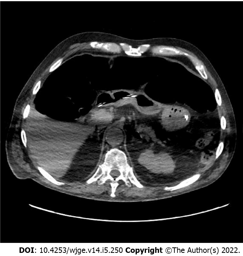
Figure 1 Case of percutaneous endoscopic gastrostomy failure.
Subsequent computed tomography scan showed colonic interposition between the stomach with nasogastric tube and the anterior abdominal wall due to fecal stasis.
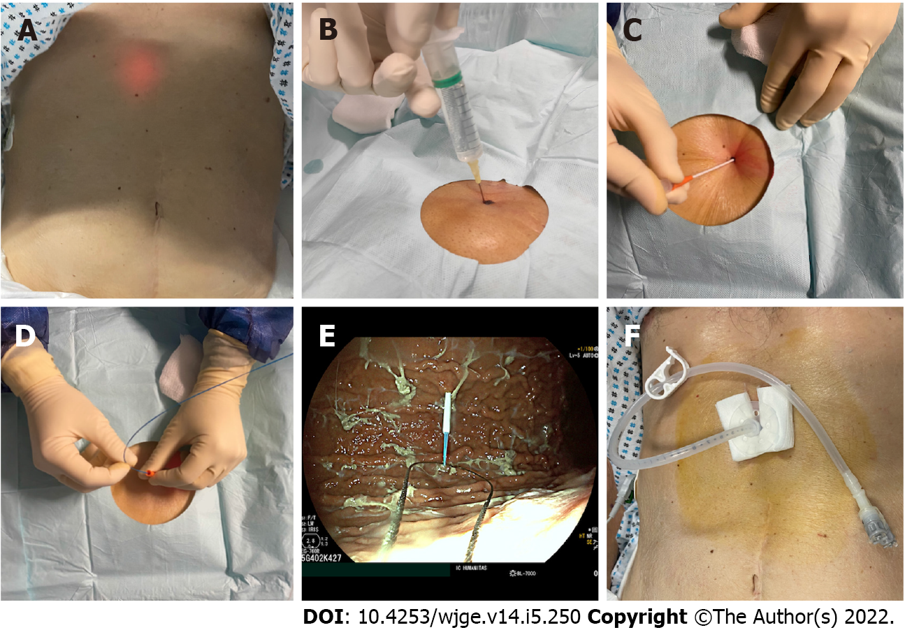
Figure 2 Steps of percutaneous endoscopic gastrostomy placement with “pull” technique.
A: Location of the puncture site via transillumination; B: Avoidance of bowel interposition confirmed by the absence of bubbles at aspiration; C: Introduction of the trocar; D: Introduction of the guidewire; E: Grasping the guidewire with an endoscopic snare; F: Final result.
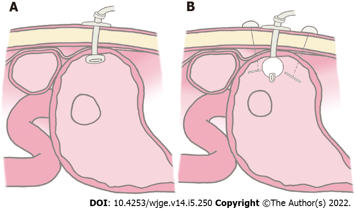
Figure 3 Graphic representation of percutaneous endoscopic gastrostomy placement technique.
A: “Pull” technique; B: “Introducer” technique.
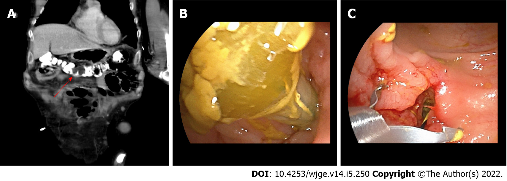
Figure 4 Percutaneous endoscopic gastrostomy displacement and development of colocutaneous fistula.
A: Computed tomography scan image showing percutaneous endoscopic gastrostomy balloon located in the transverse colon (red arrow); B: Endoscopic view of the percutaneous endoscopic gastrostomy balloon within the colon; C: Endoscopic closure of the colonic fistulous orifice with clips.
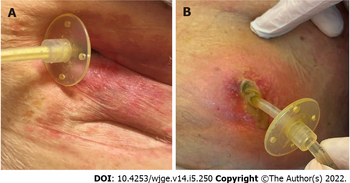
Figure 5 Wound infections.
A: Superficial infection of the abdominal wall; B: Wound infection with abscess formation within the anterior abdominal wall.
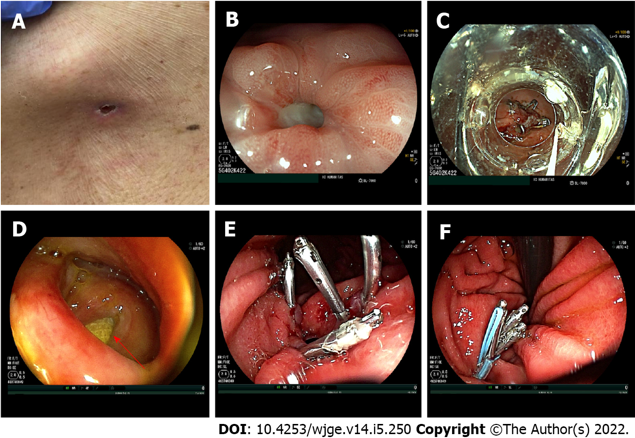
Figure 6 Gastrocutaneous fistula.
A: External appearance of a gastrocutaneous fistula in the first case; B: Endoscopic appearance of the gastrocutaneous fistulous orifice; C: Endoscopic closure of the gastric fistulous orifice with an over-the-scope metal clip in the first case (OTSC – Ovesco Endoscopy AG, Tubingen, Germany); D: Endoscopic appearance of a large gastrocutaneous fistula, with detection of the gauze placed from the outside at the cutaneous end of the tract (red arrow) in the second case; E: Endoscopic placement of four metal clips at the margins of the fistulous orifice; F: Placement of an endoloop over the metal clips to achieve complete closure of the fistulous orifice.
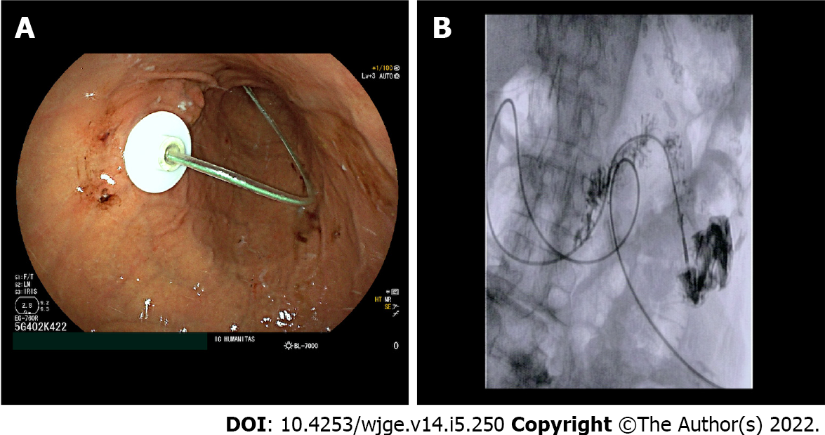
Figure 7 Percutaneous endoscopic transgastric jejunostomy placement.
A: Endoscopic appearance of the percutaneous endoscopic transgastric jejunostomy with jejunal extension entering from the percutaneous endoscopic transgastric device towards the jejunum; B: Final fluoroscopic appearance of the percutaneous endoscopic transgastric jejunostomy with distal end of the jejunal extension into the proximal jejunum after injection of contrast medium.
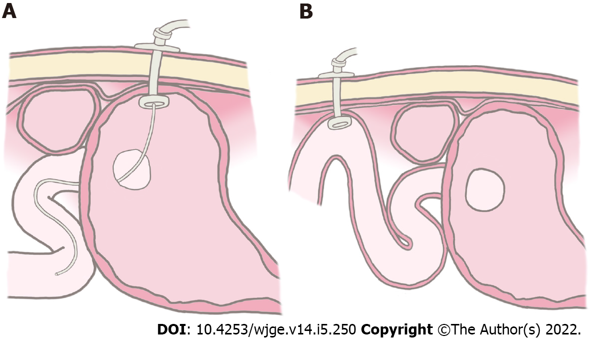
Figure 8 Graphic representation.
A: Percutaneous endoscopic gastrostomy with jejunal extension; B: Direct percutaneous endoscopic jejunostomy.
- Citation: Fugazza A, Capogreco A, Cappello A, Nicoletti R, Da Rio L, Galtieri PA, Maselli R, Carrara S, Pellegatta G, Spadaccini M, Vespa E, Colombo M, Khalaf K, Repici A, Anderloni A. Percutaneous endoscopic gastrostomy and jejunostomy: Indications and techniques. World J Gastrointest Endosc 2022; 14(5): 250-266
- URL: https://www.wjgnet.com/1948-5190/full/v14/i5/250.htm
- DOI: https://dx.doi.org/10.4253/wjge.v14.i5.250


